Santiago Ramón y Cajal
Article
Life and discoveries of Santiago Ramón y Cajal
by Marina Bentivoglio
This article was published on 20 April 1998.
Biographical sketch
Santiago Ramón y Cajal was born in May 1852 in the village of Petilla, in the region of Aragon in northeast Spain. His father was at that time the village surgeon (later on, in 1870, his father was appointed as Professor of Dissection at the University of Zaragoza). Cajal was a rebellious teenager, and his father apprenticed him for a while to a shoemaker and to a barber. Cajal, however, had decided to become an artist. His passion for drawing, his sensitivity to visual esthetics and his talent in converting visual images into drawings remained the hallmarks of his future scientific activity. Finally enrolled in the medical school at Zaragoza, as a young student, Cajal, seized by a “graphic mania,” was very fond of philosophy and gymnastics, restless, energetic, shy and solitary. He graduated in medicine at the University of Zaragoza in 1873. Shortly after his degree he was drafted into the army and dispatched to Cuba, at that time under Spanish rule, as a medical officer. Cajal returned to Spain very sick (he had contracted malaria in Cuba, and then tuberculosis), and at the end of 1875 he started his academic career as “Auxiliary Professor” of Anatomy at the University of Zaragoza.
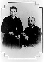
Portrait of Cajal with his wife in their first years in Madrid.
In 1879 he married Silvería Fañanás García, a non-educated young woman, who stood at his side for the rest of their lives (she died in 1930). They had seven children (two of them died in their childhood).
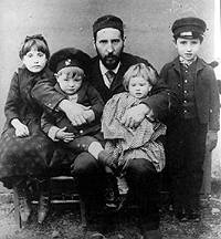
Self-portrait of Cajal with his children (from left to right: Fe, Jorge, Pula and Santiago) in Barcelona.
In Zaragoza, Cajal purchased in 1877 with his own funds (“using every peseta saved from the service in Cuba”), an old-fashioned microscope and started his scientific activity. His first studies were devoted to inflammation and to the structure of muscle fibers. In 1883, Cajal was appointed to the chair of Anatomy in Valencia. In 1885, during his tenure as Professor at the University of Valencia, the Provincial Government of Zaragoza, in recognition of his labor during a cholera epidemic, awarded him with a modern Zeiss microscope. At the end of 1887 Cajal moved to Barcelona, where he accepted the chair of Normal and Pathological Histology, and in 1892 he was appointed Professor of Histology and Pathological Anatomy at the University of Madrid. Cajal continued to work productively in Madrid until his death in 1934.

Self-portrait of Cajal in his laboratory in Valencia.
A flash of lightning
The key event for Cajal’s scientific career and for the development of modern neuroscience took place in Madrid in 1887, when Cajal was 35 years old. In this year, Luis Simarro Lacabra, a brilliant psychiatrist interested in histological research, showed to Cajal, who had traveled from Valencia to get an update on technological advances, material impregnated with the Golgi staining. Dr. Simarro had just returned from Paris, and had brought specimens stained by the new technique of silver impregnation (the reazione nera), that had been discovered 14 years earlier by Camillo Golgi but still had a very limited diffusion. Cajal wrote in his autobiography “it was there, in the house of Dr. Simarro … that for the first time I had an opportunity to admire … those famous sections of the brain impregnated by the silver method of the Savant of Pavia.”
Microscope slides with Cajal’s histological preparations; the letter ‘b’ (bueno, good) indicates the quality of the sections.
At the time, Cajal had only been studying the nervous system for one year, mainly to collect suitable illustrations for a book of histological techniques, and he had realized how inadequate the ordinary methods were to study the nervous tissue. The observation of preparations impregnated by the Golgi stain was a flash of lightning: “a look was enough” and Cajal was enraptured. Nerve cells appeared “coloured brownish black even to their finest branchlets, standing out with unsurpassable clarity upon a transparent yellow background. All was sharp as a sketch with Chinese ink,” Cajal wrote in his autobiography. In a feverish burst of activity (“… as new facts appeared in my preparations, ideas boiled up and jostled each other in my mind. A fever for publication devoured me”), Cajal worked on the retina, the cerebellum and the spinal cord, applying to the tissue the Golgi stain, of which he worked out some modifications.
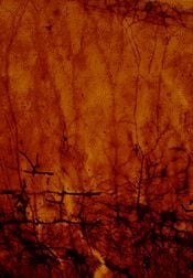
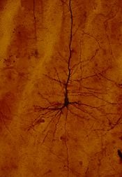
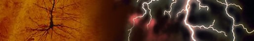
Photomicrographs from Cajal’s preparations (housed in the Museo Cajal at the Cajal Institute, Madrid, Spain) of the cerebral cortex of a newborn infant, showing neurons impregnated by the Golgi stain. The material was kindly provided by Dr. Javier DeFelipe; reproduced with the permission of Dr. Ricardo Martínez-Murillo, Director of the Cajal Institute, CSIC, Madrid. These two photos have also been published in DeFelipe and Jones “Cajal on the Cerebral Cortex.” Oxford University Press, New York, 1988.
An indefatigable and creative scholar
In October 1889, Cajal who had never traveled outside Spain except for his service in Cuba, went to Berlin, to the Congress of the German Anatomical Society, to show his slides to the leading authorities in the field, in order to convince them of the importance of his observations. On this occasion, he obtained the recognition of several qualified professors, including the eminent Swiss histologist Rudolf Albert von Kölliker (1817-1905), who from there on became a supporter of Cajal and of the “neuron doctrine,” which would be officially enunciated by Wilhelm Waldeyer (1836-1921) in 1891.
Cajal was fiercely opposed to the idea that the nervous system was made up a network of continuous elements, as it had been stated by Joseph von Gerlach (1820-1896) and supported by Golgi himself. Camillo Golgi had believed to have found in his own preparations the demonstration that the nervous system was made of a widespread network of filaments in continuity one with the other (the rete nervosa diffusa, ‘diffuse neural network’). On the contrary, since the first observations and in his subsequent studies, Cajal’s imagination was fired by the idea that the nervous system is made up of billions of separate nerve cells. Cajal’s work led to the conclusion that the basic units of the nervous system were represented by individual cellular elements (which Waldeyer christened as “neurons” in 1891). This conclusion is the modern basic principle of the organization of the nervous system.
Cajal’s opus “Textura del Sistema Nervioso del Hombre y los Vertebrados” (1894-1904), was made available to the international scientific community in its French translation, “Histologie du Système Nerveux de l’Homme et des Vertébrés”, (translated by L. Azoulay, published in 1911 by Maloine, Paris; the English translation, by N. and L.W. Swanson, was published in 1994 by Oxford University Press). Cajal’s opus provided the foundation of modern neuroanatomy, with a detailed description of nerve cell organization in the central and peripheral nervous system of many different animal species, and was illustrated by Cajal’s renowned drawings, which for decades (and even nowadays) have been reproduced in neuroscience textbooks.
Cajal’s drawing of the cerebellar cortex (from a preparation based on Golgi impregnation of a kitten cerebellum). The letter A marks the Purkinje cells with their characteristic dendritic ramifications.
Cajal’s drawing of the cerebellar cortex (from a preparation of the cat cerebellum stained with methylene blue) showing the axons of Purkinje cells which exit from the cortex directed downwards.
Preparation through the optic tectum (from a sparrow) impregnated with the Golgi technique. Note the variety of neurons drawn by Cajal.
Superficial layers of the human frontal cortex drawn by Cajal on the basis of Golgi impregnation. The main cell types of the cerebral cortex i.e. small and large pyramidal neurons (A, B, C, D, E) and non pyramidal (F, K) cells (interneurons in the modern nomenclature) are superbly outlined.
In addition, Cajal defined “the law of dynamic polarization,” stating that the nerve cells are polarized, receiving information on their cell bodies and dendrites, and conducting information to distant locations through axons, which turned out to be a basic principle of the functioning of neural connections. Cajal also made fundamental observations on the development of the nervous system and its reaction to injuries (his volume “Degeneration and Regeneration of the Nervous System” translated and edited by R. M. May, London, Oxford University Press, 1928, has been re-edited by J. DeFelipe and E.G. Jones, Oxford University Press, 1991).
Golgi and Cajal, who shared the Nobel Prize in 1906 for their studies on the nervous system, met only in Stockholm, to receive the award. Golgi gave his Nobel lecture first, in which he tied to his belief in “reticular” neural networks, which was entirely contradicted by Cajal’s Nobel lecture. Cajal, a strenuous supporter of the contiguity (and not the continuity) of individual cells representing the basic units of the nervous system, fought for his ideas until his death.
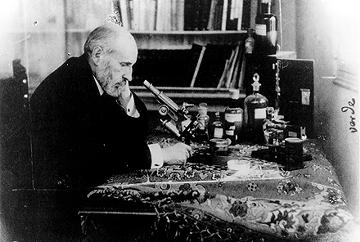
Self-portrait of Cajal at the microscope in 1920.
Golgi and Cajal certainly shared the same passion for science and dedication to science but their personalities were very different. Cajal, impetuous, burning with enthusiasm, dedicated his life to the study of the organization of the nervous system, on which he made fundamental discoveries with his peculiar talent and intuition. Golgi, a “cooler” academic, discovered the tool used by Cajal in his studies and provided outstanding contributions in many fields of cell biology and of pathology, and important contributions also on the structure of the nervous system (such, for example, the description of branches given off by the axon, of different types of neurons, of glial cells). However, Golgi misinterpreted the overall view of the structural organization of the nervous system, which instead has been worked out by Cajal.
Extremely productive, Cajal was also an accomplished photographer (his photographs of Spain, villages, friends, faces, are kept at the Cajal Museum in Madrid), and he wrote several books destined to a not strictly scientific wide audience, including his autobiography “Recollections of My Life” (Recuerdos de mi vida, translated by E.H. Craigie with the assistance of J. Cano, MIT Press, Cambridge, Mass., 1989), a small volume of aphorisms (“thoughts, anecdotes and confidences,” as stated in the subtitle) entitled “Coffee Chatters” (Charlas de Café), “The world seen at 80 years” (“El mundo visto a los ochenta años,” with the ironic subtitle of “Impresiones de un Arteriosclerótico”).
Credits
Cajal’s four drawings from “Histologie du Système Nerveux de l’Homme et des Vertébrés” were reproduced by the permission of Dr. Ricardo Martínez-Murillo, Director of the Cajal Institute, CSIC, Madrid. The portrait and self-portraits of Cajal were taken from the book “Santiago Ramón y Cajal o la Pasión de España” by Agustín Albarracín, published by Editorial Labor, S.A. (1982).
First published 20 April 1998
Nobel Prizes and laureates
Six prizes were awarded for achievements that have conferred the greatest benefit to humankind. The 14 laureates' work and discoveries range from quantum tunnelling to promoting democratic rights.
See them all presented here.

