Press release

NOBELFÖRSAMLINGEN KAROLINSKA INSTITUTET
THE NOBEL ASSEMBLY AT THE KAROLINSKA INSTITUTE
9 October 1981
The Nobel Assembly of Karolinska Institutet has today decided to award the Nobel Prize in Physiology or Medicine for 1981 with one half to
Roger W. Sperry
for his discoveries concerning “the functional specialization of the cerebral hemispheres”
and the other half jointly to
David H. Hubel and Torsten N. Wiesel
for their discoveries concerning “information processing in the visual system”.
Summary
The cerebrum is made up of two halves, the hemispheres, which are structurally identical. These hemispheres are united to one another through a system consisting of millions of nerve fibers. Therefore, each hemisphere is continually informed about what is happening in the other. For more than a century we have known that, despite their similarities and close linking, the two hemispheres generally perform different functions. The left hemisphere is the center for speech and, accordingly, has been described as the dominant one and has been considered to be superior to the right hemisphere. Outside of this, little was known about where in the brain the higher functions were centered until the beginning of the 1960s when Sperry began his investigations. Sperry has brilliantly succeeded in extracting the secrets from both hemispheres and in demonstrating that they are highly specialized and also that many higher functions are centered in the right hemisphere.
Of all the sensory impressions proceeding to the brain, the visual experiences are the dominant ones. Our perception of the world around us is based essentially on the messages that reach the brain from our eyes. For a long time it was thought that the retinal image was transmitted point by point to visual centers in the brain; the cerebral cortex was a movie screen, so to speak, upon which the image in the eye was projected. Through the discoveries of Hubel and Wiesel we now know that behind the origin of the visual perception in the brain there is a considerably more complicated course of events. By following the visual impulses along their path to the various cell layers of the optical cortex, Hubel and Wiesel have been able to demonstrate that the message about the image falling on the retina undergoes a step-wise analysis in a system of nerve cells stored in columns. In this system each cell has its specific function and is responsible for a specific detail in the pattern of the retinal image.
Roger W. Sperry
Normally, both cerebral hemispheres are linked through the cerebral commissure, which is built up of hundreds of millions of nerve fibers. When Sperry in the beginning of the 1950s began his experimental studies on animals, the functional significance of these connections between both hemispheres was entirely unknown. In experiments on monkeys Sperry found that, if these connections were severed, each cerebral hemisphere would retain its ability to learn, but that what had been learned by one hemisphere was not accessible to the other. A neurosurgical technique, so-called commissurotomy, which was similar to what Sperry had performed on monkeys, had at that time also been carried out in a number of patients suffering from severe, intractable epilepsy. A majority of these patients showed an improvement as well as a decrease in the frequency of epileptic seizures. Otherwise, the operation entailed no obvious changes at all with regard to the patients’ general behaviour and reactions. Nor could one demonstrate with psychological test methods any impairment at all in the patients’ ability to perceive and learn. When, early in the 1960s, Sperry had the opportunity to study these patients he was able, through brilliantly designed test procedures, to show that each cerebral hemisphere in these patients had its own world of consciousness and was entirely independent of the other with regard to learning and retention. Moreover, each had its own world of perceptual experience, emotions, thoughts and memory completely out of reach of the other cerebral hemisphere.
As Sperry was able to demonstrate, the isolated left hemisphere is concerned with abstract thinking, symbolic relationships and logical analysis of details, particularly temporal relationships. It can speak, write and make mathematical calculations; in its general function it is analytical and computer-like (see figure 1). It is also the more aggressive, executive, leading hemisphere in control of the nervous system. The right hemisphere is mute and generally lacks the possibility to communicate with the outside world. It is, as Sperry expresses it, “a passive, silent passenger who leaves the driving of behaviour mainly to the left hemisphere”. Because of its muteness, the right hemisphere has so far been completely inaccessible for experimental studies, and also, as a consequence of this, has been considered as being entirely subordinate to the left hemisphere. Through his investigations, Sperry has revealed that the right hemisphere, contrary to what one previously thought, is clearly superior to the left hemisphere in many respects. This is especially true regarding the capacity for concrete thinking, spatial consciousness and comprehension of complex relationships. It is also the superior hemisphere when it comes to interpreting auditory impressions and in comprehension of music; it can better recognize melodies and better distinguish voices and intonations. In other respects, however, the right hemisphere is clearly inferior to the left. It lacks almost entirely the ability to calculate and can only perform simple additions up to 20. It completely lacks the power to subtract, multiply or divide.
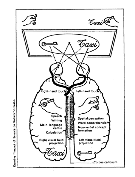 |
| Figure 1. Schematic illustration of the specialization of both cerebral hemispheres. |
It can read and comprehend the meaning of simple, mono-syllabic nouns but cannot perceive the import of adjectives or verbs. It cannot write but is entirely superior to the left hemisphere when it comes to space perception and reproducing three-dimensional pictures (see figure 2). Almost 50 years ago the great Russian physiologist Ivan Pavlov concluded that mankind can be divided into thinkers and artists. Perhaps the left hemisphere is the dominant one in thinkers and the right hemisphere in artists.
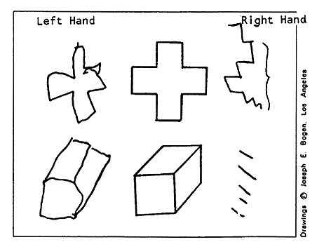
Figure 2. The ability of both hemispheres to reproduce a picture. A patient with severed connections between both his cerebral hemispheres was asked to draw the cross and cube seen in the middle of the picture. Despite the fact that he was right-handed, he was almost entirely incapable of reproducing the pictures with his right hand (which is controlled by the left hemisphere), whereas he was able to do it relatively well with his left hand (which is controlled by his right hemisphere).
In short, with his studies on commissurotomized patients, Sperry has achieved something that was previously considered almost unattainable: He has provided us with an insight into the inner world of the brain, which hitherto had been almost completely hidden from us. With his discoveries of the specialization of both cerebral hemispheres he has given us an entirely new dimension in our comprehension of the higher functions of the brain.
David H. Hubel and Torsten N. Wiesel
At the time Hubel and Wiesel began their studies of the visual system, knowledge of the functional organization of the cerebral cortex was fragmentary. By tapping nerve-cell impulses in the various layers of the visual cortex, Hubel and Wiesel have been able to demonstrate that the message reaching the brain from the eyes undergoes an analysis in which the various components of the retinal image are interpreted with respect to their contrasts, linear patterns and the movement of the image across the retina. This analysis occurs in a rigid sequence from one nerve cell to another in which each nerve cell is responsible for a certain detail in the image pattern. To put it extremely simply, one can say that the visual cortex’s analysis of the coded message from the retina proceeds as if certain cells read the simple letters in the message and compile them into syllables that are subsequently read by other cells, which, in turn, compile the syllables into words, and these are finally read by other cells that compile words into sentences that are sent to the higher centers in the brain, where the visual impression originates and the memory of the image is stored.
Hubel and Wiesel found in their studies of the visual cortex that the cells are arranged in a regular manner in columns, and that the cells within each such column have the same functions in interpreting the impulse message from the eyes. These columns make up, in turn, so-called hypercolumns, and each such hypercolumn occupies a portion of the cerebral cortex about two-by-two millimeters in area. Within each such area the information arriving from a correspondingly small region of each eye is analyzed.
Hubel and Wiesel were also able to show by their experiments that the ability of the cells in the visual cortex to interpret the code of the impulse message from the retina is developed directly after birth. A prerequisite for this development to take place is that the eye be exposed to visual stimuli. If one eye is closed for only a few days during this period, permanent functional changes will take place in the visual cortex. Hubel and Wiesel were able to show that light stimulation in itself was insufficient to bring about normal development of the visual cortex, and that it was necessary for the retinal image to have a pattern and many contours.
This discovery illustrates, first, the brain’s high degree of plasticity immediately following birth and, second, how important it is that the brain receive a rich variety of visual stimuli during this period. It is only a slight exaggeration to say that what we see today, i.e., how we perceive the visual world around, depends on the visual experiences we had during the first stages of our lives. If the visual impressions are dull or distorted – for example, through errors in the lens system of the eye – this may lead to a permanent impairment of the ability of the brain to analyze visual impressions.
The discoveries of Hubel and Wiesel represent a break-through in research into the ability of the brain to interpret the code of the impulse message from the eyes. Thanks to their investigations we now have a deeper insight into information analysis within the visual system and into the processes forming the basis for the origin of the visual impression.
Reference Material
R.W. Sperry: Split-Brain. Approach to Learning Problems. The Neurosciences. A Study Program. 1967. pp. 714. Rockefeller University Press, N.Y.
R.W. Sperry: Lateral Specialization in the Surgically Separated Hemispheres. The Neurosciences. 3rd Study Program. 1974. pp. 5. Rockefeller University Press, N.Y.
M. Gazzaniga & J. Le Doux: The Integrated Mind. Plenum Press. 1978.
D.H. Hubel: The Brain. Scientific American 1979, vol. 241, pp. 38.
D.H. Hubel & T.N. Wiesel: Brain Mechanisms of Vision. Scientific American 1979, vol. 241, pp. 130.
S.W. Kuffler & J.G. Nicholls: From Neuron to the Brain. Sinauer Assoc. Inc. Publishers 1976.
D. Ottoson: Nervsystemets Fysiologi. Natur och Kultur, Stockholm, 1978.
Nobel Prizes and laureates
Six prizes were awarded for achievements that have conferred the greatest benefit to humankind. The 14 laureates' work and discoveries range from quantum tunnelling to promoting democratic rights.
See them all presented here.

