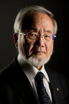
Yoshinori Ohsumi – Biographical
1. Early life and influences
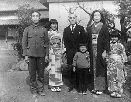
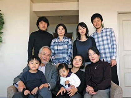
I was born in 1945 in Fukuoka, half a year before the end of the World War II, to my father Yoshio and mother Shina. My father was employed at Kyushu University as a professor of mining engineering. At that time, everyone in Japan was equally poor, and even food was not readily available. I was a weak child who continually fell ill, a condition that was perhaps exaggerated by the malnourishment of the era. My mother, after giving birth to me, contracted tuberculosis and spent a long time battling the disease in bed. I can still remember the sight of her in plaster after developing spine caries. Thankfully, a friend of my parents in Hawaii sent just-developed antibiotics which allowed my mother to recover. Words such as streptomycin and aureomycin stuck in my mind as a child, although I didn’t fully understand their meaning. When I was about 8 years old, my mother was finally able to resume a normal life. My brother, Kazuo, who is 12 years my senior, entered the Faculty of Literature at the University of Tokyo as I began elementary school. I was brought up through the efforts of my father, my two sisters Reiko and Junko and many other people who assisted through a difficult period for our family. If I had met a wonderful doctor at such a time, I might have chosen a medical education. However, I had a dream to become a scientist that was fuelled by the expectations of my parents.
I was brought up in a house surrounded by nature, with paddies, streams, hills and the sea all nearby. My youngest years were spent outdoors, catching fish and picking plants. During elementary school, I became interested in insect collecting and spent much time searching for unusual specimens. I also loved to look up at the arching night sky and remembered the names of stars and constellations. But without anyone around to foster these interests, they were left behind as childhood hobbies rather than lifelong passions. Every time my brother returned home from Tokyo during university holidays, he would bring a book, including thoughtfully chosen scientific books that broadened my awareness of the world beyond what we were taught at school. Being a weak child, I had no talent for sports and was neither artistic nor gifted in literature. But I managed to achieve good results at school.
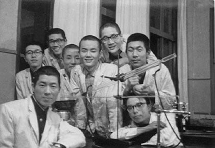
During my high school years, I was a member of the chemistry club, where we would mix chemicals and enjoy watching the reactions between them. I distinctly remember the beauty of the Liesegang phenomenon in agar, but having no idea how to analyse it. I hoped to become a chemist and entered the University of Tokyo. However, the chemistry classes did not rouse my interest and I went through an uncertain period during which I agonised over my future. Thankfully, at that time the exciting field of molecular biology was just being established, and I decided to major in it. As a graduate student, I joined the laboratory of Kazutomo Imahori, who was one of the few scientists running a molecular biology laboratory in Japan at the time. Imahori’s special field was physicochemistry to assess interactions between proteins and nucleic acids. Under the direction of Akio Maeda, I began to study the role of ribosome subunits in protein synthesis. Although I was unable to produce particularly interesting results, my time in the Imahori lab was exciting as we participated in the work of a rapidly developing field and I got my first taste of the joy of experimental work. The experience also provided me with a strong sense of the continuous nature of protein synthesis within cells.
The period when I was a graduate student was characterised by large student protests, and Tokyo University was right in the middle of the social debate and change of the period. I wasn’t able to completely focus on my lab work as I participated in the discussions and demonstrations. However, I believe that I gained a sense that we must be aware of social developments and the direction of society during this time.
From the second year of my doctoral studies, I decided to move to Kyoto University, where Maeda had joined the just-founded Department of Biophysics as an associate professor. Kyoto University provided a very free environment, and I was stimulated by talented new students and friends in biochemistry lab in the Department of Chemistry. In my research, I became interested in colicin E3, which is able to pass through the membrane of bacterial cells and instantly inhibits protein synthesis. I think I was also influenced by my growing interest in biological membranes during this period.
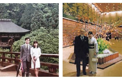
One year later, in 1971, I married Mariko Nakazawa, who joined Imahori’s laboratory two years after me. The year we spent in Kyoto after we got married was very memorable for me. However, Mariko unexpectedly became pregnant, and she therefore returned to Tokyo after less than a year. In order to support our new family, she successfully applied to the newly formed Mitsubishi Life Science Institute. I also returned to Tokyo to follow Imahori, who had moved to the Faculty of Agriculture at the University of Tokyo, and I barely managed to finish a thesis to graduate with my PhD.
At the time, it was not easy to get a good academic position. Imahori advised me that the field of cell biology was just taking off and suggested that I work in the laboratory of G. M. Edelman at The Rockefeller University. At the time, Ichiro Yahara, a senior student of Imahori’s, was playing an active part there, so I was immediately offered a position. It was my first time leaving Japan and was also a very different field to my previous work, so I felt very uncertain. Mariko decided to leave the Institute and joined me in New York. Fortunately, Mariko soon was able to find a research position at the same university, in the laboratory of Norton Zinder.
Initially I was to study mitogenesis in lymphocytes, but Edelman decided to shift the focus of the lab to mouse development, so I became involved in setting up an in vitro mouse fertilisation system. I had only ever handled Escherichia coli, so I would agonise daily over how to capture the process of early development with only a small number of eggs. Before long, Mike Jazwinski transferred to our laboratory from Arthur Kornberg‘s lab. Taking inspiration from the elegant uncovering of the cdc mutants by Hartwell, a project began to uncover the mechanism of DNA replication initiation in yeast, which I joined. We had first planned to use isolated nuclei in this project using density-gradient centrifugation, but it proved difficult to obtain undamaged nuclei. I noticed a white layer at the top of the tube, and when I looked at this layer under the microscope out of mere curiosity I realised that we were also isolating highly purified vacuoles. This left a strong impression on me and was one of those serendipitous steps that led me to where I am today.
In the autumn of 1977 Yasuhiro Anraku, a professor at Tokyo University’s Faculty of Science, asked me to return to Japan as an assistant professor in his laboratory, and while I’d just started our work on yeast DNA replication initiation, I decided to hurry back to Japan. In December 1977, I brought my soon to be 6-year-old son back to Japan, while Mariko stayed for several months longer with our recently born second son in New York to finish her work in Zinder’s laboratory.
Anraku’s laboratory was renowned for its work on transporters and the respiratory mechanism in E. coli, but Anraku allowed me to start a project on yeast. Looking back, I really appreciate Anraku’s decisiveness in affording me this opportunity. I eventually decided to study not the plasma membrane but rather the membrane of the vacuole, a cellular organelle. At the time, the vacuole was thought of as little more than a garbage dump of the cell, and there were few scientists interested in it, but my position in the Department of Botany as well as location next to Masashi Tazawa’s laboratory went some way in convincing me that the vacuole was an underappreciated, fascinating organelle. I’m sure that many yeast researchers thought it a strange choice for a project.
Soon I had developed a procedure for the isolation of vacuole membrane vesicles from highly purified vacuoles. Although the vacuole makes up approximately one quarter of the volume of the cell, I could only obtain a surprisingly small amount of vacuolar membrane, but was able to demonstrate the active transport of amino acids and calcium over the vacuolar membrane. In addition, we were the first to show the novel proton pump V-type ATPase, which gives the driving force for vacuolar transport, works that were the fruit of collaboration with the late Yoshimi Kakinuma and Etsuko Uchida. Following this, structural and functional studies of V-type ATPase became an active area of research that continues to this day.
2. Moving to Komaba and discovering autophagy
Following this decade of work on the vacuolar membrane, I was offered a place at the University of Tokyo’s College of Arts and Sciences (Komaba Campus) as an associate professor and started my own laboratory – consisting of only myself. My lab was very modestly equipped, with just a shaker, incubator, spectrophotometer, basic light microscope and a few other instruments. I realised that this was a really rare opportunity to start an entirely new subject, and at our first departmental meeting announced that I would examine the lytic function of the vacuole. However, I didn’t have any meticulously crafted strategy for how I could study this question. Since the vacuole is an acidic compartment that contains a range of lytic enzymes, I hypothesised that this organelle would play a similar role to the mammalian lysosome. The segregation of the potentially hazardous degradation process within a membrane compartment makes biological sense, but I knew at the time that if this were true material destined for degradation would have to be delivered across the vacuolar membrane, suggesting that it would require a cell biological approach and not be easy to tackle. My work on protein degradation originates from this research on the vacuole; I was 43 years old.
The lysosome was discovered by Christian de Duve in 1955, and soon after researchers at The Rockefeller University observed the delivery of both extracellular materials and cytoplasmic materials to the lysosome by electron microscopy. De Duve coined the term autophagy to describe the latter process, from the Greek for ‘self-eating’. Although some, most notably Glenn Mortimore and Per Seglen, had conducted intricate analyses of the regulation and complex membrane dynamics of autophagy, there were many more questions than answers when I joined the field. The complexity of lysosome and membrane dynamics in mammalian cells prevented the uncovering of the autophagy mechanism. De Duve’s lab was actually in the same building as Edelman’s, and while I had friends from his lab, I never had the chance to meet with this great scientist at The Rockefeller University.
Light microscopes today, including the development of fluorescence microscopy, are advanced instruments, but at that time the power of light microscopy was limited and little information about the intracellular events could be obtained, especially for the tiny yeast cell. However, the yeast vacuole could be clearly observed under phase contrast optics. The inside of the vacuole is somewhat like a salt solution with a very low protein concentration, so I knew that it would be easy to detect structures within the vacuole due to the difference in refractive index. As I would observe the vacuole every day at the microscope, I think I probably spent longer watching yeast cells than anyone else during these years!
First of all, I searched for a condition where yeast would break down their own proteins and settled on sporulation as the best candidate. This dramatic change, from vegetatively growing cell to four spores via meiosis, is induced by the environmental depletion of nitrogen. As nitrogen is key ingredient of protein, I reasoned that the protein synthesis required for such drastic cellular remodelling must depend on the turnover of the cell’s own proteins. I therefore closely observed the morphology of the vacuole during sporulation, but wasn’t able to observe any obvious changes.
One day, I conceived that if I were to use a cell lacking vacuolar proteases, structures delivered to the vacuole for degradation would remain in this organelle and might be visible. Fortunately, Elizabeth Jones had already constructed strains lacking vacuolar proteases and, in an example of the sharing ethos of the yeast community, had deposited them into the Yeast Genetic Stock Center at UC Berkeley, so I immediately requested that they be sent to my laboratory. When they arrived, I subjected them to nitrogen starvation and examined them under the microscope. I observed small, spherical structures moving vigorously within the vacuole. These structures appeared within 30 minutes of the onset of nitrogen starvation and had completely filled the vacuole within a few hours. In the otherwise tranquil yeast cell, this dramatic phenotype really struck me, and I distinctly remember staring down the microscope for hours, unable to tear my gaze away. These structures are immediately degraded in wild-type cells, which is why they had never been properly documented. I was fortunate for two reasons: first, the structures inside the vacuole, which we now call autophagic bodies, were big enough to see under the microscope; and second, the intense movement of autophagic bodies by Brownian motion made their accumulation in the vacuole easy to notice, even with a basic light microscope. The observation of this phenomenon determined the course of the rest of my career.
I decided to immediately determine what causes this phenomenon by electron microscopy and enlisted the help of Misuzu Baba and Masako Osumi. The images they captured were overwhelmingly beautiful and provided unequivocal evidence of the membranous phenomena characteristic of autophagy in yeast. These images showed that autophagic bodies contain the same density of ribosomes as the cytoplasm as well as various cytoplasmic structures and organelles, suggesting that autophagy is a non-selective degradative process. These experiments provided us with our current understanding of the membrane dynamics associated with autophagy. Although the vacuole is vastly larger than the mammalian lysosome, the entire process that we observed was topologically the same as macroautophagy in mammalian cells, confirming that yeast could be used as a model organism in the study of autophagy.
The most attractive feature of yeast as a model organism is its genetic tractability, lending it great power in the unpicking of complex biological phenomena. Two important examples that strongly influenced me were Lee Hartwell’s identification of the cdc mutants and their function in the cell cycle, and Randy Schekman’s isolation of the sec mutants and their role in membrane trafficking.
As we knew nothing of the genes and proteins that are involved in the autophagy, the first step was therefore to isolate autophagy-defective mutants. But the only means we had of monitoring autophagy was to observe autophagic body accumulation in the vacuole. My first Master’s degree student, Miki Tsukada, was central at this stage of our work. She took thousands of mutagenised cells, individually subjected them to starvation and then checked for mutants lacking autophagic bodies in the vacuole. This approach led us to identify the first autophagy mutant strain, which we called apg1. A failure to induce protein degradation in this strain convinced us that autophagic bodies are indeed responsible for degradation during starvation. As expected, we also found that the homozygous diploid mutant was unable to sporulate. However, under nutrient rich conditions no difference was observed between the apg1 strain and wild-type cells, and there appeared to be no obvious defect in vacuolar function or secretion.
Soon we found that in comparison to their wild-type counterparts, apg1 cells rapidly lose viability during nitrogen starvation. Assuming that this phenotype was caused by a defect in autophagy, we next conducted a preliminary screen assessing viability. By isolating strains in which viability was reduced and then examining these strains for morphological defects in autophagy, we were able to identify roughly 100 autophagy-defective strains in a single screen. Further genetic analysis showed that 14 complementation groups were among these strains. Looking from our current understanding of autophagy, it’s clear that this screen was extremely effective. We initially predicted that these mutants would be characterised by defects at various stages of the autophagy process, but surprisingly all were essential for the formation of the autophagosome, the most important step in autophagy.
Next, we set out to clone and identify the genes implicated in the mutant phenotypes observed in the screen. Tsukada first succeeded in the cloning of the APG1 gene, through which we learned that this gene encodes a protein kinase. However, we also found that the remaining genes encoded uncharacterised proteins essential for autophagy, but not required for growth under rich conditions. Therefore, we were unable to deduce anything about their function and went through a difficult period where we were unable to publish our exciting findings. Although we made rapid progress, autophagy genes had somehow escaped the attention of other researchers in spite of the comprehensive genetic analyses of yeast all around the world. I suspect that this is because researchers were interested in essential genes, conventionally defined by inviability in nutrient rich medium. At this time, Takeshi Noda also developed a quantitative assay of autophagic activity, the still widely-used Pho8Li60 assay. These developments together laid the foundations for our subsequent studies on the molecular mechanism of autophagy.
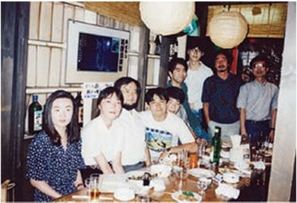
This is how my career in autophagy research began in that small laboratory at the University of Tokyo. The whole process, from uncovering autophagy to beginning the characterisation of the Atg genes, was achieved within eight years in Komaba.
NIBB
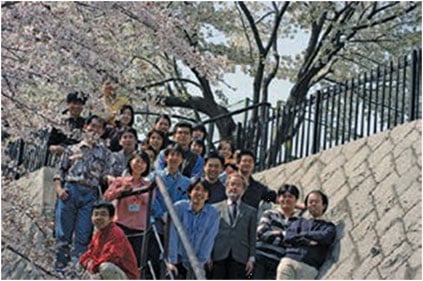
The next phase began in 1996, when I was chosen as a professor at the National Institute for Basic Biology (NIBB) in Okazaki. This facility provided an environment very supportive of basic biology, and I selected three researchers to join our team: Tamotsu Yoshimori, originally of Kansai Medical School, who would work to incorporate our findings in yeast into mammalian cells; Takeshi Noda, who was the first doctoral student to graduate from our lab at Komaba; and Yoshiaki Kamada, who had studied at Johns Hopkins University in the US. The following year, Noboru Mizushima, previously a clinician at Tokyo Medical and Dental University, also joined the laboratory as a JSPS postdoctoral researcher and later as an assistant professor. From this point, graduate students and postdocs from all over Japan joined our lab and we entered the most fruitful age of our research. At the NIBB, we were able to produce a truly unique research environment where we extended our work to include mammalian cells and plant cells, which was important in the development of the field. Most members of our laboratory lived near the NIBB, and as both Yoshimori and I were working away from home during this period we would often work late into the night, discussing science over a beer.
I was expecting that it would take a long time to isolate all the genes required for autophagy, but thanks to our collaboration with my wife Mariko’s laboratory, which helped with cloning at the Teikyo University of Science, as well as the sequencing of the entire yeast genome in 1996, we were able to elucidate the genetic context of nearly all the APG genes in a relatively short period.
After that, we undertook to identify interacting proteins of the characterised Apg proteins, and were able to confidently identify all 18 of the genes essential for autophagy under nitrogen starvation conditions. A group led by Michael Thumm was studying the reduction of protein levels using antibodies under starvation conditions and was close behind our group, naming genes they identified using the ‘aut‘ alias. Meanwhile, Dan Klionsky’s group was investigating the delivery of aminopeptidase Ape1 by cytosol to vacuole targeting, now known as the Cvt pathway, identifying genes using the ‘cvt‘ alias. This pathway is in fact a biosynthetic pathway responsible for the maturation of Ape1. Through the collaboration of our groups, we determined that this was occurring through similar membranous phenomena that depend on the core autophagic machinery and very selectively isolate Ape1 for delivery to the vacuole. Klionsky’s group thus uncovered the Cvt pathway, which served as an important model of what was to become the major field of selective autophagy.
With the continued isolation of autophagy-defective mutant strains in yeast by many groups, we collectively decided to unify gene names under the ATG alias to simplify nomenclature in the field. As the majority of the key autophagyrelated genes were identified by our group, the gene numbers used by our group when assigning APG gene names was retained in this system. With the identification of further autophagy-related genes, most of which are involved in selective autophagy, the number of ATG genes has now reached 41.
During our time at the NIBB, important findings came one after the next as we began to look at the ATG gene products to gain insights into their function. Mizushima made the first important breakthrough in his investigation of the only very recently cloned Atg12 protein. He discovered an intricate conjugation system that functions in an ubiquitin-like manner to yield a dimer of Atg12-Atg5-Atg16 complexes essential for autophagy. Two graduate students, Takayoshi Kirisako and Yoshinobu Ichimura, also uncovered another ubiquitinlike conjugation system that, unusually for such a system, results in the conjugation of Atg8 with a membrane phospholipid, phosphatidylethanolamine. Atg8, homologues of which were soon identified in mammals by Yoshimori and in plants by our group, remains associated with the autophagosome throughout all stages of autophagy and is therefore widely used as a marker of autophagy progression. We therefore learned that nearly half of the essential Atg proteins were involved in just two conjugation reactions. Important structural details of the complexes involved in these reactions were solved thanks to our close collaboration with Fuyuhiko Inagaki and Nobuo Noda’s groups.
Further details came with the identification of the autophagy-specific PI3 kinase complex, which we termed PI3 kinase complex I. Akio Kihara’s work showed that Atg6 had in fact only just been identified as Vps30, a protein of the vacuole protein sorting pathway, by Scott Emr’s group. Vps30 is a component of the PI3 kinase complex, and we subsequently found that Atg14 affords this complex its specific role in autophagy. Yet another specific component of PI3 kinase complex I, Atg38, was uncovered more recently by Yasuhiro Araki. Meanwhile, we also discovered the important role of Atg13 in the activation of Atg1 kinase function, and identified Atg17, Atg29 and Atg31 as forming a complex required for autophagy induction under starvation conditions. With our investigations of Atg9, Atg18 and Atg2, we were before long able to classify all 18 genes essential for autophagy under starvation conditions into six functional groups. This was further explored by Kuninori Suzuki, who used fluorescence microscopy to discover the pre-autophagosomal structure (PAS), a dynamic structure to which all Atg proteins at least partially assemble and intricately co-ordinates autophagosome formation. Suzuki’s work also demonstrated that the Atg proteins associate with the PAS in a hierarchical manner to perform their essential role in autophagy, shedding further light on the mechanisms of autophagy.
Identification of the ATG genes has completely revolutionised autophagy research. One important example of this was Mizushima’s demonstration of the progression of autophagy in mammals by using a GFP-tagged LC3 transgenic mouse, as well as his successful generation of an autophagy knockout mouse, the first for Atg5. Along with the knockout of Atg7 in mice by Masaaki Komatsu, these studies changed our understanding of autophagy in mammalian cells and spurred the widespread genetic manipulation of ATGgenes in various cells, tissues, organs and whole organisms all over the world. This movement helped to uncover a variety of physiological functions of autophagy. Of these, the recycling function of autophagy is the most fundamental in the cell, helping to survive the commonly-encountered challenge of nutrient limitation. But autophagy is not only important for nutrient recycling: it is also implicated in the often selective purging of unnecessary or harmful intracellular components, which is important in the maintenance of the intracellular environment. Recently, the selective elimination of proteins, supramolecular structures, and organelles have come to the fore as physiologically important applications of the core autophagy machinery. A particularly salient example of this has been the intensely-studied field of selective mitochondrial autophagy, or mitophagy. The removal of old and potentially damaged mitochondria by autophagy ensures efficient energy production and a healthy intracellular environment, underscoring the nuanced integration of autophagy into the cell.
4. Starting my final lab at the Tokyo Institute of Technology
Even in an organism as apparently simple as the yeast cell, many mysteries remain unresolved. My lab has therefore been working exclusively in yeast for 13 years since Yoshimori and Mizushima formed their own groups.
In 2009, another important change occurred in my career. The Tokyo Institute of Technology offered me a position as a specially appointed professor in a laboratory with excellent facilities, so I decided to return to the Tokyo area. With Hitoshi Nakatogawa and Suzuki as assistant professors, and later Hayashi Yamomoto, we started a new laboratory. I brought along many colleagues from Okazaki to this new facility. We have been focussed one subject, how the autophagosome is formed, through the functional analysis of Atg proteins. Recently we found how the early steps of PAS formation are organised. Another area of interest to us was the mechanism of selective autophagy in yeast. In this field, we were also able to identify various receptors for target selection. Hitoshi Nakatogawa identified novel receptors for ER and nucleus degradation.
Now, my lab has shifted its focus to the physiological meanings of autophagy. In order to return to the original questions that intrigued me at the start of my career – the what, when, how and why of autophagic degradation – we need to systematically consider cells as they exist in an array of environmental contexts. This year we have set up a research unit at the Tokyo Institute of Technology, and with Tomoko Kawamata as an assistant professor we are now running a laboratory consisting mainly of postdoctoral researchers that will be my last laboratory. Here we are tackling those original questions that I believe are important to deepen our understanding of autophagy. Yeast is still an extremely powerful model organism, and with the continued development of new biochemical techniques I believe that we still have many ground-breaking contributions to make, especially in identifying the induction conditions and various modes of autophagy. These contributions will necessarily assess the products that arise from autophagic degradation, the mechanism of their export from the vacuole and their implications for cellular metabolism. As a fundamental cellular process, autophagy is linked directly and indirectly to a range of cellular pathways, and the development of quantitative methods that will allow us to grasp the subtle characteristics of this mechanism are essential. A better understanding of autophagy promises to help characterise the ambiguous relationship between genetics, phenotype and disease.
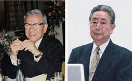
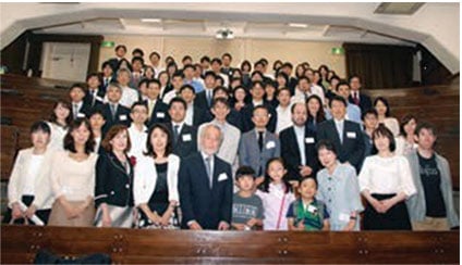
Autophagy research is still at an early stage, and our understanding of the physiological role of autophagy in particular is only in its infancy. Of course, I’m very pleased that we have managed to unveil such intricate detail about autophagy and its associated phenotypes, and the attention the field has attracted from around the world is very exciting. But I am most appreciative for the many wonderful colleagues I have worked with over the years. I was always driven by curiosity rather than immediate practical outcomes in my work, and have been lucky to work with postdoctoral researchers and graduate students who appreciate the importance of basic research. Many of them have shared my passion for studying difficult projects that do not always give immediate outcomes, and fortunately many of them continue to advance the field at a range of universities as researchers. I have also been very fortunate to have worked with many collaborators over the years who have helped us along the way. In particular I want to note the contribution of two researchers who have collaborated closely with us for over a decade in the systematic structural analysis of the Atg proteins: Nobuo Noda, who works now at the Institute of Microbial Chemistry, and Fuyuhiko Inagaki, who regretfully passed away in 2016. I deeply appreciate their collaboration over the years.
I am deeply moved by the interest that has been shown in autophagy research, which for me has represented an incredible journey over the last 28 years. This was only possible thanks to grant support of our fundamental research, which was indispensable in this undertaking. It is my greatest pleasure and honour as a basic scientist if our work was able to trigger a development in our understanding of life. I await the continued development of the field over the years to come with great anticipation.
This autobiography/biography was written at the time of the award and later published in the book series Les Prix Nobel/ Nobel Lectures/The Nobel Prizes. The information is sometimes updated with an addendum submitted by the Laureate.
Yoshinori Ohsumi – Nobel Lecture
Yoshinori Ohsumi delivered his Nobel Lecture on 7 December 2016 at Aula Medica, Karolinska Institutet in Stockholm. He was introduced by Professor Maria Masucci of the Nobel Assembly.
Autophagy – an Intracellular Recycling System: Lecture slides
Pdf 34 MB
Copyright ©: Yoshinori Ohsumi
Read the Nobel Lecture
Pdf 3.2 MB
Yoshinori Ohsumi – Nobel diploma
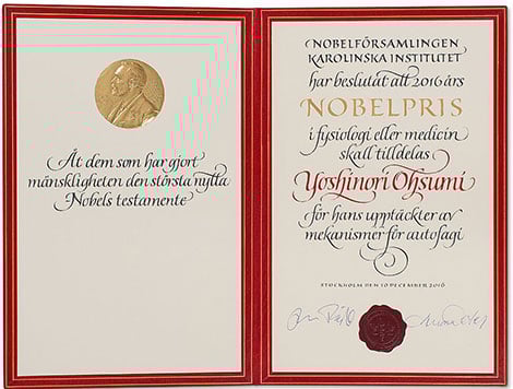
Calligrapher: Susan Duvnäs
Book binder: Leonard Gustafssons Bokbinderi AB
Photo reproduction: Lovisa Engblom
Copyright © The Nobel Foundation 2016
Yoshinori Ohsumi – Photo gallery
Yoshinori Ohsumi receiving his Nobel Prize from H.M. King Carl XVI Gustaf of Sweden at the Stockholm Concert Hall, 10 December 2016. © Nobel Media AB 2016. Photo: Pi Frisk
Yoshinori Ohsumi after receiving his Nobel Prize at the Stockholm Concert Hall, 10 December 2016.
Copyright © Nobel Media AB 2016
Overview from the Nobel Prize Award Ceremony at the Stockholm Concert Hall, 10 December 2016. © Nobel Media AB 2016. Photo: Alexander Mahmoud
Nobel Laureates at the Nobel Prize Award Ceremony. Far right: Medicine Laureate Yoshinori Ohsumi. From left: Chemistry Laureates Jean-Pierre Sauvage, Sir J. Fraser Stoddart and Bernard L. Feringa. Far right: Medicine Laureate Yoshinori Ohsumi.
Copyright © Nobel Media AB 2016
Yoshinori Ohsumi and Catharina Lindqvist at the table of honour at the Nobel Banquet, 10 December 2016.
© Nobel Media AB 2016. Photo: Alexander Mahmoud
Yoshinori Ohsumi delivering his banquet speech.
Copyright © Nobel Media AB 2016
Yoshinori Ohsumi takes a look at his Nobel diploma during his visit to the Nobel Foundation on 12 December 2016.
Copyright © Nobel Media AB 2016
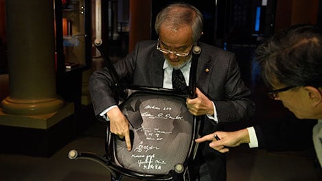
Yoshinori Ohsumi signs the Nobel Foundation's guest book, signed by the Laureates since 1952, during his visit to the Nobel Foundation. On this occasion, the Laureates retrieve the Nobel diploma and Medal, which have been displayed in the Golden Hall of the City Hall following the Nobel Prize Award Ceremony. The Laureates also discuss the details concerning the transfer of their prize money.
Copyright © Nobel Media AB 2016
Yoshinori Ohsumi showing his Nobel Medal.
Copyright © Nobel Media AB 2016
Nine of the eleven Nobel Laureates of 2016 assembled at the Nobel Foundation in Stockholm on 12 December 2016. From left: Jean-Pierre Sauvage, J. Michael Kosterlitz, David J. Thouless, Oliver Hart, F. Duncan M. Haldane, Bengt Holmström, Bernard L. Feringa, Yoshinori Ohsumi and Sir J. Fraser Stoddart.
Copyright © Nobel Media AB 2016
Yoshinori Ohsumi arriving at the Nobel Museum in Stockholm, Sweden, on 8 December 2016.
Photo: Henrik Montgomery/TT.
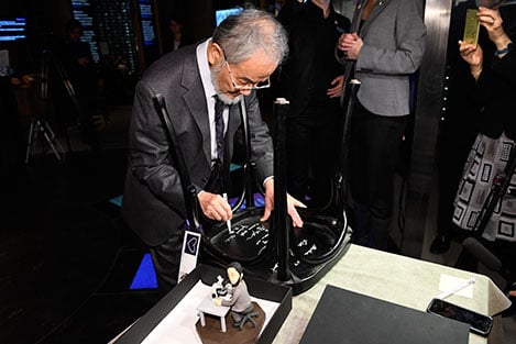
Like many Nobel Laureates before him, Yoshinori Ohsumi autographs a chair at the Nobel Museum in Stockholm.
Photo: Henrik Montgomery/TT.
Nobel Laureate Yoshinori Ohsumi and the autographed chair at the Nobel Museum in Stockholm.
Photo: Henrik Montgomery/TT.
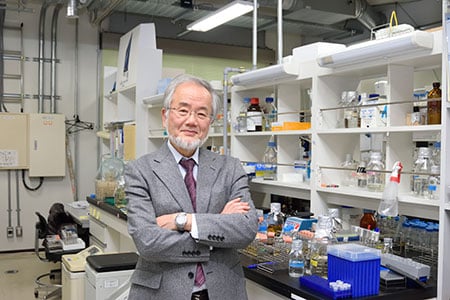
Yoshinori Ohsumi in his laboratory.
Photo: Copyright © Tokyo Institute of Technology
Courtesy: Tokyo Institute of Technology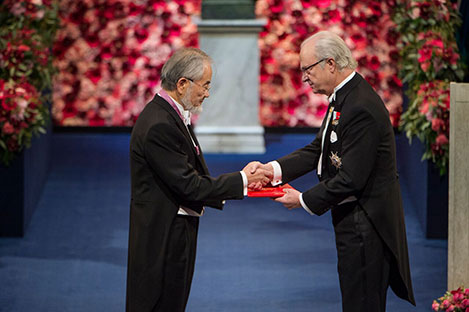
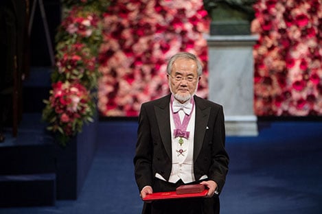
Photo: Pi Frisk 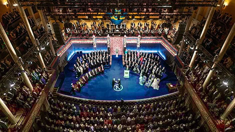

Photo: Pi Frisk 

Photo: Dan Lepp
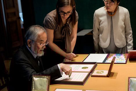
Photo: Alexander Mahmoud 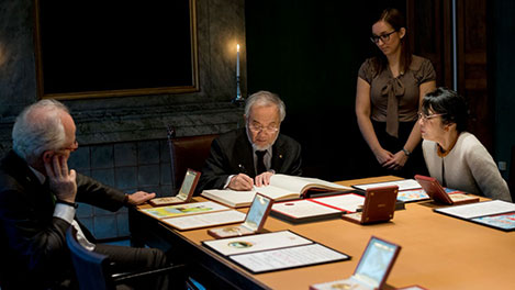
Photo: Alexander Mahmoud 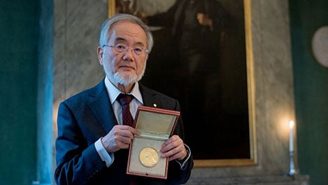
Photo: Alexander Mahmoud 
Photo: Alexander Mahmoud 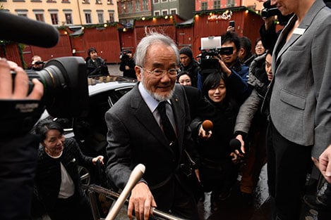
Yoshinori Ohsumi – Other resources
Links to other sites
Popular information
![]()
Populärvetenskaplig information: Cellens livsnödvändiga återvinningssystem
Swedish, pdf 1.5 MB
Yoshinori Ohsumi – Banquet speech
Yoshinori Ohsumi’s speech at the Nobel Banquet in the Stockholm City Hall, 10 December 2016.
Your Majesties,
Your Royal Highnesses,
Excellences,
Dear Laureates,
Ladies and gentlemen,
I wish to thank to the Nobel Assembly at Karolinska Institute and Nobel Foundation for awarding me the most prestigious prize in science, the Nobel Prize, in the category of Physiology or Medicine. I’d also like to congratulate this year’s other recipients. It has been lovely meeting everyone during this very enjoyable week and it is an honour to stand among such esteemed people.
I am just a basic cell biologist who has been working with yeast for almost 40 years. I would like to take this opportunity to note my appreciation for the many lessons and wonderful gifts from yeast – perhaps my favourite of all being sake and liquor.
My research career focussed on autophagy, which is a major process of recycling and degradation of proteins within cells. Life is maintained by a delicate balance between continuous synthesis and degradation. I found that degradation is just as important as synthesis for the maintenance of dynamic biological systems like the body.
My group’s contribution was to find the molecular underpinnings of autophagy. Autophagy is now exploding into one of the most intensely studied topics in biology. While our contribution is fundamental, it is pleasing that many researchers are now studying its relevance in health and trying to conquer a range of diseases. There is no greater satisfaction as a scientist than seeing your ideas and efforts transform a field of research, and I am as happy as I could be. I will finish by acknowledging the fortune, large number of excellent collaborators, indispensable grant support and caring family that brought me here tonight. Thank you again all for this wonderful opportunity.
Yoshinori Ohsumi – Interview
Interview, December 2016
Interview with Chemistry Laureate Yoshinori Ohsumi on 6 December 2016 during the Nobel Week in Stockholm, Sweden. The interviewer was freelance journalist Lotta Fredholm.
Interview, October 2016
“Still we have so many questions.”
Telephone interview with Yoshinori Ohsumi following the announcement of the 2016 Nobel Prize in Physiology or Medicine, 3 October 2016. The interviewer is Adam Smith, Chief Scientific Officer of Nobel Media.
Interview transcript
[Yoshinori Ohsumi]: Moshi, Moshi, Yoshinori speaking.
[Adam Smith]: Hello, Professor Ohsumi. My name is Adam Smith. I’m calling from the official website for the Nobel Prize.
YO: Ah ha, yeah.
AS: First of all, our congratulations on the award of the Nobel Prize.
YO: Thank you so much. Yes, I was surprised.
AS: [Laughs] How did you hear the news?
YO: I had a call from Thomas Perlmann.
AS: Yes, Secretary of the Nobel Committee, yes indeed.
YO: Yeah, Nobel Assembly.
AS: Nobel Assembly, yes. And where were you when you received the news?
YO: I was in my lab.
AS: And your first reaction?
YO: I heard that, single only, me! It was also a surprise for me.
AS: It’s true, because it’s rare that they give the Nobel Prize in Physiology or Medicine to just a single Laureate. What do you think this says about the role of the single researcher these days?
YO: That’s my real surprise because so many people are now working in the autophagy field.
AS: Autophagy is a huge area. But it’s not…
YO: Yeah, recently.
AS: Recently, exactly.
YO: Just recently I think.
AS: Very largely because of your work.
YO: Yes, it’s so, developed fast, yeah. When I started my work, probably every year 20 or less papers appeared on autophagy. Now more than 5,000 or something like that. It’s a huge change within probably these 15 years or so.
AS: A real explosion.
YO: I actually started more than 27 years ago.
AS: It was a good choice of field.
YO: Yeah, it was lucky. Yeah, yeast was a very good system and autophagy was a very good topic to work. Still we have so many questions. Even now we have more questions than when I started.
AS: Once again it underlines the power of yeast as an experimental model.
YO: Yeah.
AS: You can do so much with yeast. And does it surprise you how similar yeast is to ourselves?
YO: I believe there are fundamental functions of the cells should be conserved from yeast to mammals. So that’s my belief. But of course vacuole is different from lysosome, but I thought that most fundamental mechanisms must be conserved. That was my assumption when I started my work.
AS: You sound as if you’re still in a slight state of shock.
YO: Yeah, mmm.
AS: And will you be coming to Stockholm in December to receive your award?
YO: Yeah, yeah.
AS: Wonderful. Well we very much look forward to meeting you then and to talking further.
YO: OK.
AS: Thank you very much indeed for speaking to us now.
YO: Thanks so much.
AS: Congratulations again and we wish you a wonderful day.
Did you find any typos in this text? We would appreciate your assistance in identifying any errors and to let us know. Thank you for taking the time to report the errors by sending us an e-mail.
The Nobel Prize Award Ceremony 2016
Watch the Nobel Prize Award Ceremony from the Stockholm Concert Hall in Sweden, 10 December 2016.
Opening Address by Professor Carl-Henrik Heldin, Chairman of the Board of the Nobel Foundation
Presentation speech for the Physics Prize
Presentation speech for the Chemistry Prize
Presentation speech for the Medicine Prize
Presentation speech for the Literature Prize
Presentation speech for the Prize in Economic Sciences
Yoshinori Ohsumi – Prize presentation
Watch a video clip of the 2016 Nobel Laureate in Physiology or Medicine, Yoshinori Ohsumi, receiving his Nobel Prize medal and diploma during the Nobel Prize Award Ceremony at the Concert Hall in Stockholm, Sweden, on 10 December 2016.