Perspectives: With a little help from my friends
Godfrey Hounsfield’s groundbreaking concept of viewing organs from outside the body was so ambitious that it would require the most successful pop band in history and visionary doctors to help his idea reach fruition.
Few bands can rightly claim to have had an impact on the history of music, and even less can claim to have had a similar impact on the history of medicine. But The Beatles were a band that constantly pushed the boundaries of innovation in popular music, so it is no surprise to discover that their success helped fuel other advances – in this case the development of one of the most important diagnostic tools in the field of medicine.
Around the time the Beatles were finishing what many consider their most innovative album, Sergeant Pepper’s Lonely Heart’s Club Band, an electrical engineer working at Electric and Musical Industries (EMI) called Godfrey Hounsfield was on a weekend ramble in the English countryside, where he conceived a wondrous but ambitious idea. In fact Hounsfield’s idea for viewing and examining organs from outside the body was so ambitious it would require considerable financial investment to get it off the ground.
Thanks to The Beatles, however, whose record sales had almost doubled EMI’s profits since they had signed to its Parlophone label five years earlier, EMI had begun to invest a sizeable amount of money into funding bold research ideas. Within the space of five years Hounsfield’s idea would come to fruition, and few medical achievements would be received with such unreserved enthusiasm as would his invention of computed tomography (CT).
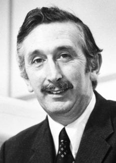
Hounsfield joined EMI in 1951, where he initially worked on radar and guided weapons. Among his achievements, his development of Britain’s first all-transistor computer, EMIDEC 1100, in 1958, stood out. EMI moved Hounsfield to its Central Research Laboratories after selling off its computer division in 1962, and assigned him to the project of designing a one-million-word immediate-access thin-film computer memory. This project failed for commercial reasons, but trusting Hounsfield’s creative capabilities, his supervisors gave him free rein to choose his next research project.
It was at this point that Hounsfield had his flash of inspiration on his countryside walk. Hounsfield was thinking about his radar research, in particular the problems of pattern recognition. Radar systems scan their surroundings by sending out radio waves from a central point and detecting patterns in the periphery. Why not try the reverse process, Hounsfield thought while walking, and study the central or interior pattern of an object from outside? Why not send beams through a parcel to find out what’s hidden inside?
“I thought, wouldn’t it be nice if I had many readings taken from all angles through a box,” said Hounsfield. “Wouldn’t it be nice if I could reconstruct in 3-D what was actually in the box from these random direction readings taken through the box?” The trick, thought Hounsfield, was to view the three-dimensional object as a series of cross-sectional scans, or slices, and he started working out mathematically how he could do this.
Hounsfield thought that X-rays could fulfil this purpose. X-rays are a phenomenally powerful tool for diagnosing fractures of bones. However, X-rays are less useful in diagnosing conditions affecting soft tissues like the brain, as the rays can’t distinguish one type of tissue from another – so they appear in X-ray photographs as a grey fog-like mass.
Hounsfield’s idea centred on the fact that the intensity of an X-ray beam reduces when it passes through an object – a process called attenuation. Different parts of the human body – for instance, bones and soft tissues like the brain – dampen X-rays differently. If you could observe the different attenuation patterns from a human object by directing an X-ray beam through it from different angles, hypothesized Hounsfield, you could distinguish between different types of tissues and reconstruct an image of a ‘slice’ of that object.
Hounsfield sent his research proposal to EMI under the title “An improved form of X-radiography”, in which he proposed that a series of X-ray exposures taken from different angles around an area of the body could construct a cross-sectional image of a ‘slice’ of that area. The different X-ray exposures could be detected by a sensing device that was always pointing towards the source of the gamma rays, and these readings would be digitized and fed to a computer to build up a crude picture of the material within the ‘slice’.
Hounsfield was unaware that the Austrian mathematician Johann Radon and the South African physicist Allan Cormack had already shown in theory that such an image could be obtained. For instance, Cormack devised a mathematical solution to measuring the tissue-density distribution within the body. On the basis of this, he proposed that X-rays could be taken from different angles around the brain or body, and accounting for the different effects of soft and dense tissues on X-rays, a computer could assemble these images into three-dimensional representations, but he did not take this idea further in terms of creating an instrument that could carry this out.
However, thanks to EMI’s deep pockets Hounsfield could begin to turn his idea into a working product. The project created enough excitement that the British Department of Health got involved through its Radiological Advisor Evan Lennon.
Hounsfield’s first experimental system used gamma rays from the radioactive element Americium to scan bottles or perspex jars filled with water and pieces of metal and plastic, and was “very much improvised”, as he recalled in his Nobel Lecture. A lathe bed provided the means for moving and rotating the gamma-ray source, and sensitive detectors were placed on either side of the bottles or jars. The scanning process took nine days and created 28,000 measurements, which took a high-speed computer two and a half hours to calculate and process. The images the computer created, though, were good enough to convince both EMI and the Department of Health to invest £6,000 each in the acquisition of an X-ray tube and a generator, which would reduce scanning time to nine hours. Hounsfield travelled across London by underground bringing bullock’s and pig’s brains fresh from the abattoir to his laboratory, and he produced the first pictures in which white and grey matter could be clearly differentiated.
Clinical Champion
The images from animal brains showed that the method worked, but Hounsfield needed to collaborate with a clinician to show whether it would work on human brains. The Department of Health’s Lennon tried in vein to find such a contact, but he came up against a wall of scepticism. “Why should I meet such a crank?” responded the first radiologist Lennon approached, and several more refused a request to meet Hounsfield.
Lennon persisted and eventually struck gold with a consultant radiologist called Jamie Ambrose, who was based at the radiology department of Atkinson Morley’s Hospital in Wimbledon, London. Ambrose was exploring ways of imaging the living brain using methods such as ultrasound and echo encephalography, and he agreed to meet Hounsfield. The first meeting did not go well. Ambrose found Hounsfield a difficult character; a man who was not very talkative, wary of explaining any details of his invention, and who responded to all the newest neurological images that Ambrose showed him with a dismissive “I can do better than that.”
As they made their goodbyes after what seemed an unfruitful meeting Ambrose handed Hounsfield a jar containing a brain with a tumour and asked him for some proof of his invention. When Hounsfield returned he brought a picture of the brain with him, which showed the tumour and even areas of bleeding within the tumour. Ambrose was instantly stunned.
Ambrose’s foresight and enthusiasm would prove to be a much-needed fillip for Hounsfield and his invention. Radiologists by and large were looking for ways in which to improve the resolution of X-ray images and reduce the time taken to get them. A new and revolutionary technique promising images of the brain, but that had less resolution and that took longer to acquire than existing methodologies wasn’t top of the wish list for most radiologists – or so they thought.
The prototype of what was called the EMI brain scanner was installed at Atkinson’s Morley Hospital and the first human patient was examined on October 1, 1971. (The EMI brain scanner was later renamed computed tomography, and the method is also known as computerized axial tomography, or CAT.) For the first CT scan Ambrose chose a woman in her early forties with a suspected brain tumour.
“There was a beautiful picture of a circular cyst right in the middle of the frontal lobe,” Hounsfield recalled, “and, of course, it excited everyone in the hospital who knew about this project.” After seeing the image Hounsfield and Ambrose felt, as the latter recalled, like footballers who had just scored the winning goal. The skull was no longer inaccessible from outside, the intricacies of the brain were now visible and they could distinguish between healthy and diseased tissue. Over the next few weeks they confirmed the capabilities of the scanner with around ten other patients, in whom they diagnosed and localized their brain diseases and thus made them accessible for surgical intervention.
When Ambrose presented these first clinical images at the Annual Congress of the British Institute of Radiology – the oldest radiological society in the world – on 20 April 1972 the audience was stunned. Seeing images of the brain that clearly showed lesions, tumours and haemorrhage instantly blew away all the scepticism radiologists had about the technique. These powerful images convinced radiologists that they were witnessing a new era in the detection and evaluation of disease.
The Department of Health purchased the first three EMI scanners produced and placed them in the Manchester Royal Infirmary, in Glasgow and at the Institute of Neurology in London. Two more scanners were sent to the Mayo Clinic and the Massachusetts General Hospital in the United States. In October 1972, Jamie Ambrose displayed and presented images from an EMI scanner to an audience of 2,000 medical doctors at the Chicago meeting of the Radiological Society of North America. He received a standing ovation, one of many that he and Hounsfield would receive over the next few years, as improvements in CT allowed sections of the body to be analysed, and the radiology literature began to reveal the full impact of the technique on the diagnosis and treatment of patients.
The invention of computed tomography provides an almost textbook example of how the progress of science relies as much on the belief and championing of ideas as it does on the quality of the idea itself. As one historian in the field says the risky realization of computer tomography could have happened nowhere else than in the United Kingdom at this particular time: “It is hard to imagine how the instrument would have gone into production without the support of a company like EMI. The combination of the Beatles’ success with the British system of research subsidies and the genius of one engineer broke the cash barrier and changed the face of modern medicine.”
Bibliography
Dixon, Adrian K.: Computed Tomography. Encyclopaedia of Life Sciences, 2001.
Hounsfield, Godfrey N.: Computed Medical Imaging. Nobel Lecture, December 8, 1979.
Husband, J. and Dombrowe, G. X-ray computed tomography – a truly remarkable medical development. British Journal of Radiology 78, 97–98 (2005).
Obituary Jamie Ambrose: The Guardian, May 10, 2006.
Obituaries Sir Godfrey Hounsfield: Independent, August 20, 2004; Telegraph, August 16, 2004; British Medical Journal 2004; 329; 687 (18 September); European Radiology 2004; 14; 2152–2153).
Petrik, Vladimir et al.: Godfrey Hounsfield and the dawn of computed tomography. Neurosurgery 58: 780–787 (2006).
Wells, P.N.T.: Sir Godfrey Newbold Hounsfield KT CBE. Biogr. Mems Fell. R. Soc. 51, 221–235 (2005).
Speed read: Clearing the fog
The famous X-ray photograph Wilhem Röntgen took of his wife’s hand showed both the potential and the limitations of using X-ray images in medicine. The bones of Röntgen’s wife’s hand can be clearly seen, as can her wedding ring, but soft tissues, blood vessels and nerves are all invisible. Over 70 years later, computed tomography, commonly known as CT or CAT scans, has taken X-ray imaging to another level by integrating X-rays with digital technology to generate three-dimensional views of inner organs and soft tissues.
Allan Cormack devised a mathematical method for measuring different tissue densities within the body, and he predicted that these calculations could be used to create X-ray images of cross-sectional slices of organs like the brain. Godfrey Hounsfield brought Cormack’s predictions to fruition, by developing a method of his own that collects a series of X-ray exposures taken around an area of the body to construct an image of a ‘slice’ of that area, and by constructing the first clinically usable machine that could create these images.
Like Röntgen’s X-ray images, Hounsfield’s first published computed tomography images in 1972 stunned the world. Up until that time, X-ray images of the head clearly showed the skull bones, but the brain looked like a grey, undifferentiated fog. With computed tomography, the fog suddenly cleared. Doctors could now see clear images of cross-sections of the brain, with the grey and white matter and liquid-filled cavities clearly visible.
Computed tomography received such immediate acceptance and met with such unreserved enthusiasm that it soon became an established method for the examination of all organ systems of the body. For opening up this new avenue in medical diagnostics, Cormack and Hounsfield received the Nobel Prize in Physiology or Medicine in 1979.
Allan M. Cormack – Nobel Lecture
Allan M. Cormack held his Nobel Lecture on 8 December 1979, at Karolinska Institutet, Stockholm. He was presented by Professor Ulf Rudhe, member of the Nobel Committee for Physiology or Medicine.
Read the Nobel Lecture
Pdf 771 kB
Godfrey N. Hounsfield – Nobel Lecture
Godfrey N. Hounsfield held his Nobel Lecture on 8 December 1979, at Karolinska Institutet, Stockholm. He was presented by Professor Ulf Rudhe, member of the Nobel Committee for Physiology or Medicine.
Godfrey N. Hounsfield held his Nobel Lecture on 8 December 1979, at Karolinska Institutet, Stockholm. He was presented by Professor Ulf Rudhe, member of the Nobel Committee for Physiology or Medicine.
Read the Nobel Lecture
Pdf 1.25 MB
Allan M. Cormack – Banquet speech
Allan M. Cormack’s speech at the Nobel Banquet, December 10, 1979
Your Majesties, Your Royal Highnesses, Ladies and Gentlemen,
Godfrey Hounsfield has asked me to speak for both of us. We would most respectfully request Your Majesty to convey to the Nobel Foundation and the Nobel Assembly of Karolinska Institutet our intense gratitude for the honour which they have done us by awarding us the Nobel Prize for medicine and physiology.
There is irony in this award, since neither Hounsfield nor I is a physician. In fact it is not much of an exaggeration to say that what Hounsfield and I know about medicine and physiology could be written on a small prescription form!
While there is irony in the award, there is also hope that even in these days of increasing specialization there is a unity in the human experience, a unity clearly known to Alfred Nobel by the broad spectrum of his awards. I think that he would have been pleased to know that an engineer and a physicist, each in his own way, have contributed just a little to the advancement of medicine.
Allan M. Cormack – Other resources
Links to other sites
Godfrey N. Hounsfield – Biographical
I was born and brought up near a village in Nottinghamshire and in my childhood enjoyed the freedom of the rather isolated country life. After the first world war, my father had bought a small farm, which became a marvellous playground for his five children. My two brothers and two sisters were all older than I and, as they naturally pursued their own more adult interests, this gave me the advantage of not being expected to join in, so I could go off and follow my own inclinations.
The farm offered an infinite variety of ways to do this. At a very early age I became intrigued by all the mechanical and electrical gadgets which even then could be found on a farm; the threshing machines, the binders, the generators. But the period between my eleventh and eighteenth years remains the most vivid in my memory because this was the time of my first attempts at experimentation, which might never have been made had I lived in a city. In a village there are few distractions and no pressures to join in at a ball game or go to the cinema, and I was free to follow the trail of any interesting idea that came my way. I constructed electrical recording machines; I made hazardous investigations of the principles of flight, launching myself from the tops of haystacks with a home-made glider; I almost blew myself up during exciting experiments using water-filled tar barrels and acetylene to see how high they could be waterjet propelled. It may now be a trick of the memory but I am sure that on one occasion I managed to get one to an altitude of 1000 feet!
During this time I was learning the hard way many fundamentals in reasoning. This was all at the expense of my schooling at Magnus Grammar School in Newark, where they tried hard to educate me but where I responded only to physics and mathematics with any ease and moderate enthusiasm.
Aeroplanes interested me and at the outbreak of the second world war I joined the RAF as a volunteer reservist. I took the opportunity of studying the books which the RAF made available for Radio Mechanics and looked forward to an interesting course in Radio. After sitting a trade test I was immediately taken on as a Radar Mechanic Instructor and moved to the then RAF-occupied Royal College of Science in South Kensington and later to Cranwell Radar School. At Cranwell, in my spare time, I sat and passed the City and Guilds examination in Radio Communications. While there I also occupied myself in building large-screen oscilloscope and demonstration equipment as aids to instruction, for which I was awarded the Certificate of Merit.
It was very fortunate for me that, during this time, my work was appreciated by Air Vice-Marshal Cassidy. He was responsible for my obtaining a grant after the war which enabled me to attend Faraday House Electrical Engineering College in London, where I received a diploma.
I joined the staff of EMI in Middlesex in 1951, where I worked for a while on radar and guided weapons and later ran a small design laboratory. During this time I became particularly interested in computers, which were then in their infancy. It was interesting, pioneering work at that time: drums and tape decks had to be designed from scratch. The core store was a relatively new idea which was the subject of considerable experiment. The stores had to be designed and then plain-threaded by hand (causing a few frightful tangles on occasions). Starting in about 1958 I led a design team building the first all-transistor computer to be constructed in Britain, the EMIDEC 1100. In those days the transistor, the OC72, was a relatively slow device, much slower than valves which were then used in most computers. However, I was able to overcome this problem by driving the transistor with a magnetic core. This increased the speed of the machine so that it compared with that of valve computers and brought about the use of transistors in computing earlier than had been anticipated. Twenty-four large installations were sold before increases in the speed of transistors rendered this method obsolete.
When this work finished I transferred to EMI Central Research Laboratories, also at Hayes. My first project there was hardly covered in glory: I set out to design a one-million word immediate access thin-film computer store. The problem was that after a time it was evident that this would not be commercially viable. The project was therefore abandoned and, rather than being immediately assigned to another task I was given the opportunity to go away quietly and think of other areas of research which I thought might be fruitful. One of the suggestions I put forward was connected with automatic pattern recognition and it was while exploring various aspects of pattern recognition and their potential, in 1967, that the idea occurred to me which was eventually to become the EMI-Scanner and the technique of computed tomography.
The steps in my work between this initial idea and its realisation in the first clinical brain-scanner have already been well documented. As might be expected, the programme involved many frustrations, occasional awareness of achievement when particular technical hurdles were overcome, and some amusing incidents, not least the experiences of travelling across London by public transport carrying bullock’s brains for use in evaluation of an experimental scanner rig in the Laboratories.
After the initial experimental work, the designing and building of four original clinical prototypes and the development of five progressively more sophisticated prototypes of brain and whole body scanner (three of which went into production) kept me fully occupied until 1976. Since then I have been able to broaden my interest in a number of projects which are currently in hand in the Laboratories, including further possible advances in CT technology and in related fields of diagnostic imaging, such as nuclear magnetic resonance.
As a bachelor, I have been able to devote a great deal of time to my general interest in science which more recently has included physics and biology. A great deal of my adult life has centred on my work, and only recently did I bother to establish a permanent residence. Apart from my work, my greatest pleasures have been mainly out-of-doors, and although I no longer ski I greatly enjoy walking in the mountains and leading country rambles. I am fond of music, whether light or classical, and play the piano in a self-taught way. In company I enjoy lively way-out discussions.
This autobiography/biography was written at the time of the award and later published in the book series Les Prix Nobel/ Nobel Lectures/The Nobel Prizes. The information is sometimes updated with an addendum submitted by the Laureate.
Godfrey N. Hounsfield died on August 12, 2004.
Allan M. Cormack – Facts
Godfrey N. Hounsfield – Facts
Press release

NOBELFÖRSAMLINGEN KAROLINSKA INSTITUTET
THE NOBEL ASSEMBLY AT THE KAROLINSKA INSTITUTE
11 October 1979
The Nobel Assembly of Karolinska Institutet has decided today to award the Nobel Prize in Physiology or Medicine for 1979 jointly to
Allan M Cormack and Godfrey Newbold Hounsfield
for the “development of computer assisted tomography”.
An X-ray examination usually implies the passage of X-rays through an organ with a resulting image of the organ on X-ray film. The dark areas on the film vary according to the anatomy and the structure of the tissues being X-rayed.
A peculiarity of this picture is that it is two-dimensional. In the reproduction the dimension of depth is lost. This means that an overall picture of the lungs, for example, is a composite one in which all the details in the path of the rays are overlapped. In order to acquire any depth perception, one must complement frontal exposures with lateral exposures. The radiologist’s interpretation of possible changes in the lungs is based on his knowledge of the normal anatomy of the lungs and of the properties of the pathological abnormalities. But the nature of the final X-ray image makes judgement in certain cases undeniably subjective. Therefore, in many situations there is a need to be able to isolate the image of a section of an organ from the overlying structures by so-called tomography (from the Greek tomos, a cut, and graph, written). Many technical solutions have been tested during the course of the years but none have been found to be entirely satisfying. For purely physical reasons one can never achieve a complete eradication of other sections of the organ, and the picture’s contrast is reduced. This is true even when one allows the radiation beam to run parallel to the examined section so that the rays proceed from one edge to another. There are other limitations to conventional radiological diagnostics. One is that X-rays cannot be utilized to more than 25 %; another the X-ray film has a relatively low sensitivity in the reproduction of the variations in tissue density.
In computer-assisted tomography these problems have been ingeniously solved. When the method was introduced into medical care six years ago it quickly became apparent that it signified something revolutionarily new, with great repercussions with X-ray diagnostics and the medical disciplines that make use of it.
The basic feature of the method is that the X-ray tube, in a definite pattern of movement, permits the rays to sweep in many directions through a cross-section of the body or the organ being examined. The X-ray film is replaced by sensitive crystal detectors, and the signals emitted by amplifiers when the detectors are struck by rays are stored and analyzed mathematically in a computer. The computer is programmed to rapidly reconstruct an image of the examined cross-section by solving a large number of equations including a corresponding number of unknowns. The image presented on the screen of the oscilloscope is drawn in a fine system of squares, a so-called matrix, in which each individual square corresponds to a part of the examined organ. Each element expresses the permeability of X-rays of the corresponding part of the organ. A fundamental peculiarity is that the image elements do not influence each other while the image is being reconstructed. In other words, there is no overlapping of elements in the image. Because the sensitivity of the crystal detectors and amplifier is more than 100 times as great as X-ray film, computed tomography can detect very subtle variations of tissue density. This means that the density resolution is exceptionally high. For all practical purposes one achieves a correct image of a thin section of organ tissue.
The first computer tomograph was constructed to be used for examining the skull, with special emphasis on diseases of the brain. The method soon experienced an enormous breakthrough in the radiological diagnosis of neural diseases. The reason is the precision and sensitivity of computed tomography. Extensive special examinations, such as contrast encephalography and pneumaencephalography, that is, X-ray examination with a contrast medium in the vessels, and filling the brain cavities with air following lumbar puncture, provide very valuable information, but nevertheless indirectly. The need for these types of encephalography is now reduced. Computed tomography, on the other hand, provides in each section a very detailed picture of the brain and its cavities as well as the fluid-filled spaces surrounding the brain, i.e., the cisterns and subarachnoid spaces, everything visible directly on the picture. This means that pathological changes in the brain and its surroundings can be well demonstrated by the computed tomogram. Their position, size and shape can be estimated and their nature can often be determined. The number of black gradations in the squared pattern of the image is greater than an observer can perceive, but can be extracted from the image and denoted numerically. This appreciably simplifies determination of the nature of the disease. Hemorrhages, and such changes in the brain arising from a blood clot blocking circulation, cause the same symptoms but can be distinguished by computed tomography. Tumors and conditions brought on by inflammation, senile changes in the brain, hydrocephalus and malformations in the brain can all be revealed. The method is invaluable in developing new methods for operating on brain tumors. So rich is the detail that the computed tomogram is reminiscent of the picture one gets of the brain at autopsy.
Computer-assisted tomography cause no discomfort to the patient, who lies comfortably on his back during the examination. This makes it possible to examine even very sick individuals in an acute phase of their illness. The effect of the treatment can be monitored. All centers in the world with access to a computed tomograph attest to the fact that the method has meant an enormous advance in diagnostics, therapy, development and research within the specialty of neurological diseases.
With modern computed tomographs it is possible to examine every organ in the body. In certain connections the method is superior to all other methods. In other situations it complements other techniques, such as ultrasound, isotope diagnostics with the gamma camera.
A very important area of application, which is rapidly growing in importance, is the radioactive treatment of tumors. Heretofore the weakest link in planning radiation treatment has been the difficulty in determining, with desired precision, the position, size and shape of tumors in the innermost regions of the body. This involves the problem of delimiting tumors from surrounding tissue. With computed tomography it is possible to carefully analyze all these factors, from a scaled image of the body on a level with the tumor. This facilitates the choice of suitable radiation field and optimal ray quality. When the tumor shrinks during treatment, which can be shown by computed tomography, the radiation can gradually be changed so that more resistant sections of the tumor can be irradiated more intensely than surrounding tissue. Well-informed observers believe that computer-assisted tomography has introduced a new era in radiation therapy. The entire field is the subject of intensive research.
This year’s Nobel Prize in physiology or medicine has been awarded to Allan M Cormack and Godfrey N Hounsfield for their contributions toward the development of computer-assisted tomography, a revolutionary radiological method, particularly for the investigation of diseases of the nervous system.
Allan Cormack is professor and head of the institution of physics at Tufts University in Medford, Massachusetts, USA. He was the first, from a theoretical point of view, to analyze the conditions for demonstrating a correct radiographic cross-section in a biological system. He published his analysis of the problem in two scientific publications in 1963 and 1964. He understood that the problem was basically a mathematical one. It was a matter of finding a reasonable two-dimensional function that described how X-rays attenuate in each individual part within a slice when one knows the mean values of the rays’ absorption, the so-called line integrals, along a number of straight lines within this slice. He was convinced that the problem had great principle interest and foresaw that, if it could be solved, there would be possible applications within medicine, such as radiotherapy and positron-camera diagnostics. He was not aware then that the key mathematical problems had been considered earlier in an altogether different connection and deduced his own method of calculation. In extensive model experiments, in which he used gamma radiation that has a shorter wave-length than X-rays, he showed that the agreement between theory and experiment was good. Cormack’s reconstruction mathematics is one of several possible ones that can be used. His contributions to the development of the theory of computer-assisted tomography was early and anticipated the coming development by several years by being the first to state the principles for reconstructing a cross-section of tissues in an organ based on these X-ray projections. The reason Cormack’s discovery did not come to be industrially applied is not known, but it can be assumed that the computers of the time lacked sufficient capacity to enable the method to be applicable to medical care.
Godfrey Hounsfield, who is chief of the medical research division of Electric and Musical Industries, Middlesex, England, is the central figure in computer-assisted tomography. He has made the really decisive contributions for introducing computed tomography in medicine by constructing the first computed tomography system practicable in medical care. Thus, he described a complete system for computed tomography in his patent application in 1968. The patent was granted in 1972. An advance communication about the method came in 1971, a more extensive report with a supplement of clinical viewpoints by Ambrose followed in 1972, and a detailed description of the system appeared in the December, 1973, issue of the British Journal of Radiology. This work and the patent papers are epoch-making in medical radiology. The achievement is no less significant because all the components forming the basis for the construction and operation of the computed tomograph had been described earlier in non-medical publications. Hounsfield was obviously unaware of Cormack’s contributions and developed his own method for reconstruction of the image. With an unusual combination of vision, intuition and imagination, and with an extraordinarily sure eye for the optimal choice of physical factors in a system that must have offered very great problems to construct, he obtained results which in one blow surprised the medical world. It can be no exaggeration to maintain that no other method within X-ray diagnostics has, during such a short period of time, led to such remarkable advances, with regard to research and number of applications, as computer-assisted tomography.
Hounsfield’s system, which was directed at examinations of the skull and brain, started a development which in a few years led to the so-called fourth generation of computed tomographs. In these, technical improvements and more rapid analytical reconstruction methods have raised performance still farther, work in which Hounsfield has taken active part.