Werner Forssmann – Nobel Lecture
Nobel Lecture, December 11, 1956
The Role of Heart Catheterization and Angiocardiography in the Development of Modern Medicine
The ancient world and the Middle Ages had no idea of the existence of the circulation of the blood. It was not until the Late Renaissance that efforts were made to grasp this process anatomically and understand its function. Thus, Miguel Serveto searched in vain for a connection between the right heart and the left, and in so doing discovered the lesser circulation in 1553. In 1569, Caesalpinus traced the path of the large circulation. Jacobus Sylvius (1543), Canani (1564), and Fabricius of Aquapendente (1574) concurred in recognizing the centripetal movement of the venous bloodstream from the structure and arrangement of valves in the veins. Before their time it had been believed that blood flowed outwards to the periphery, even in the veins.
William Harvey, one of the most gifted pupils of Fabricius (1578-1657), combined all these individual findings with the results of his own research to form the general picture of what we today call the circulation of the blood. But even he had no clear idea of the circulation in the region of the capillaries. This section was explained and described for the first time by Malpighi in 1661, after he had viewed a frog’s lung under a microscope.
In any event, it is the year 1628, in which Harvey published his classic work De motu cordis et sangunis, that we can call the birth-year of cardiology.
However, this great discovery appears to have had no immediate effect on the method of observing the function of the sound and sick heart. This had to wait some 170 years, until, at the end of the eighteenth and the beginning of the nineteenth centuries, scientific methods of examination made their appearance in medicine. The beginning of this epoch was marked by the introduction of digitalis for the treatment of oedema, achieved by William Withering in 1785.
Further milestones on the way were the introduction of percussion by Auenbrugger in 1761 and of auscultation, by Laëmec. These innovations made the increasingly refined discoveries of the new science of pathology useful at the sickbed. The last landmark of this period was Einthoven’s introduction of the electrocardiogram into clinical practice and research rays, discovered shortly before, enabled the grosser modifications of the heart’s structure to be seen even in a living person.
With this, cardiology had entered a stage of stagnation, which made it necessary to seek new and more exact methods than those hitherto available.
The starting-point of the modern trend in research came from classic French experimental physiology, notably the trials on animals to obtain blood for metabolism experiments described by Claude Bernard in his Physiologie opérative. In particular, the procedure employed by Chauveau and Marey in 1861 became the model. They were the first to achieve measurement of blood pressure inside the heart and the recording of pressure curves from the interior of the heart of a living animal. This was done with manometers, which were led from the neck vessels into both compartments of the right heart as well as into the left heart chamber.
But even Claude Bernard, Chauveau, and Marey had been forestalled. As far as I know, the credit for carrying out the first catheterization of the heart of a living animal for a definite experimental purpose is due to an English parson, the Reverend Stephen Hales. This scientifically interested layman undertook in Tordington in 1710, 53 years after the death of William Harvey (1578-1657), the first precise definition of the capacity of a heart. He bled a sheep to death and then led a gun-barrel from the neck vessels into the still-beating heart. Through this, he filled the hollow chambers with molten wax and then measured from the resultant cast the volume of the heart-beat and the minute-volume of the heart, which he calculated from the pulse-beat. Besides this, Stephen Hales was also the first, in 1727, to determine arterial blood pressure, when he measured the rise in a column of blood in a glass tube bound into an artery.
In 1912, Unger, Bleichröder, and Loeb published a work under the title Intra-arterial therapy. They were at that time aiming at a special chemotherapy for puerperal sepsis. In order to bring a drug in the greatest possible concentration to the place where it was needed, they wanted to insert ureter catheters into human patients from the leg arteries up to the presumed height of the fork of the aorta and inject from there. After experiments on animals, they carried out vein probings on four people as a preliminary trial from the point of view of intra-arterial therapy. These caused no ill effects. No X-ray checks were used, nor were the cardiological aspects taken into consideration.
In 1928, the Italian Montanari carried out probing of the right heart on animals and on the human cadaver.
Here I may as well review my own first attempts at probing the right heart, undertaken in 1929 and based on the work of Chauveau and Marey.
The reason they at first made no headway was because, in the years 1929-1931, all the technical requirements for the planned investigations were lacking and had first to be laboriously created. So it can be understood that it was some time before the broad outlines of important problems of developing modern cardiology could be seen emerging from my admittedly rather unusual experimental procedure. Perhaps it is significant that the pioneer work of O. Frank and Broemser on the development of the manometer also took place in these years.
Thus the experimental methods and the results they yielded needed many years to come to fruition. They achieved their modern practical significance only because fundamental discoveries had been made in other fields, for example in modern anaesthetic techniques, in antibiotics and through the pioneering publications of Helen Taussig, which in time bore further fruit. Nevertheless, in 1930, about six months after my first publication, O. Klein reported from Nonnrenbruch’s Prague clinic on a series of patients whose heart minute-volumes he had ascertained according to Fick’s principle, by means of the heart catheter. This procedure has its place even today in the standard practice of heart and lung clinics.
At the same time; I carried out my first experiments in angiocardiography. Here for the first time the living heart of a dog was successfully visualized radiologically with the aid of a contrast medium. Even at that time, the complete lesser circulation in the dog could be shown with the cinematographic radioscopy according to Gottheiner.
Although no results could be attempted with human beings, because no apparatus had been devised, their possibility had at least been demonstrated in principle. Only four months after this publication, Moniz, Carvalho, and Lima were able to disclose rather better results. with them began the immense quantity of writing on angiocardiography.
Further development of technique was impeded not only by the absence of technical essentials and consequent lack of knowledge. To some outsiders, ethical considerations also weighed heavily in the balance against it. And when one thinks how hard men like Cournand and McMichael had to fight against such people in 1941 and later, one can perhaps understand what difficulties stood in my way twelve years before.
A turning-point in the history of cardiology is the year 1941, when Cournand and Ranges made known their first experiments with the heart catheter as a clinical method of investigation. But he will be reporting on this himself.
The work of Cournand and Richards and their pupils had fanned a small flame into a blazing fire which began to rage all over the world. The Cournand-Richards school achieved particularly fruitful results in the United States and Scandinavia. In England, McMichael is the most important advocate of this method of investigation. His great service is his employment of it to solve pharmacological problems.
For Cournand and McMichael, too, as they have told me themselves, the beginning was not easy. They, too, had strong resistance to overcome, the harder to deal with because people did not hesitate to obstruct practical research work with threadbare ethical and moral objections, such as are still occasionally raised today. But these voices also must fall silent now it has been shown, how responsibly this circulation research has been conducted everywhere and with what high moral earnestness it has been applied. And so now there is an army of diligent men and women at work, an army so big that it is impossible in this context to mention any beyond those named. You must pardon this omission.
As for angiocardiography, this method has given strong new impetus not only to cardiology, but also to X-ray technology as a whole. Whereas earlier X-ray diagnosis stopped short at explaining the morphology from the reproduction of shadows, here a leap was made right to the core of the function.
At this point, the close and inseparable interrelation of heart and lungs became obvious – something we had certainly guessed at, but which previously we could not grasp. And so, with the selective angiography of the lung vessels (Bolt, 1949-1950), our knowledge was consciously extended to the outermost periphery.
With this, heart catheterization had burst the bounds of cardiology in the stricter sense, and now set about conquering other fields of research.
Right at the start of my experiments upon animals it had occurred to me, as to other investigators, that one could penetrate diagonally through the right auricle from the upper into the lower vena cava. Now use is made of this path to collect blood from the liver, and it is to be hoped that many long-standing questions of metabolism will be solved in this way. The kidney, too, is accessible in the same manner. Thus it can be seen that heart catheterization must in no way be looked upon as an almost worked-out field of research.
Angiocardiography, in the form in which it is practised today, is of course still burdened with risks which impose limitations on its use. Its use cannot therefore be justified for examinations which are not strictly necessary, but here, too, new possibilities can be discerned.
Further development will in many cases enable us to dispense with the massive and dangerous quantities of contrast media which at the moment we still need, and to manage instead with smaller, less harmful amounts of radioactive isotopes. Their progress through the small circulation can already be followed in outline with Gripping’s isotope retina, and shown graphically.
From all this, we can see that modern cardiology has become something much more universal than was originally supposed.
One may compare the art of healing with a work of art, which from different standpoints and under different lighting reveals ever new and surprising beauty.
So, besides the epochs of cellular and humoral pathology and many others, we can now perhaps speak of an age of cardiological and circulatory investigation. We do this with the comforting awareness that, by the correct application of their teaching, earlier discoveries remain useful to us, for they now appear in a new light. Thus we guard ourselves against the mistake which runs all through the history of medicine: that of concentrating dogmatically upon first one, then another facet of research, instead of standing back to view the whole as a growing entity.
Dickinson W. Richards – Nobel Lecture
Nobel Lecture, December 11, 1956
The Contributions of Right Heart Catheterization to Physiology and Medicine, with Some Observations on the Physiopathology of Pulmonary Heart Disease
The study of the right heart in man has held a continuing place in the researches of Dr. Cournand and myself over the past twenty-five years, both under physiological and pathological conditions. Measurements made in this exact location have provided a key to almost all the syntheses, all the integrations that we have attempted, in elucidating the nature of cardiopulmonary function.
In the realm of pathology, pulmonary heart disease also occupies a key position, affected as it is by all manner of pulmonary as well as circulatory dysfunctions. With many of these we are particularly concerned in our research studies of the present day.
In expressing my acknowledgement and appreciation to the Nobel Prize Committee and to the Caroline Institute for the supreme honour which they have so generously conferred on our work, I should like now, using these two subjects as a central theme, to give a brief account of our research from its beginning.
The origins of any systematic research are many, extending widely, as well as far back in time. Many also are the supporting and sustaining research activities which are in progress simultaneously. We find ourselves deeply indebted to our colleagues throughout the world, for such assistance over many years.
The foundation upon which the work of Dr. Cournand and myself chiefly rests is that of Lawrence J. Henderson of Harvard. Biochemist, physiologist, natural philosopher, student of Arrhenius, ardent disciple of Claude Bernard, Henderson achieved as his great single contribution the definitive integration of the respiratory function of the blood1. But his horizon was far wider than this. He was a general physiologist in the broadest sense. For him the physical chemistry of the blood was but a single link in the whole circulorespiratory synthesis. Such breadth of view was inherent in everything he thought or did, and this could not help but be reflected, in some degree, in those whose good fortune it was to be associated with him. It was from Henderson that we derived the simple but essential concept that lungs, heart, and circulation should be thought of as one single apparatus for the transfer of respiratory gases between outside atmosphere and working tissues.
This concept was of course not new, was in fact in the great tradition of Krogh, of Lindhard2, of Liljestrand3,4 and their collaborators, who with the methods then available, worked out to a remarkable extent in man the performance of the normal cardiorespiratory apparatus, in rest and exercise. Liljestrand made also a number of studies in disease. In the late nineteen-twenties and early thirties, the active research team at the Harvard Fatigue Laboratory under Henderson continued this exploration of cardiocirculatory patterns1, to which their complete studies of the respiratory function of the blood constituted an important contribution.
During all this time, however, and indeed for a total period of nearly forty years, there was in the study of the heart and circulation in man, one measurement or set of measurements which were conspicuously in default, viz. the state of the blood as it enters the right heart, its respiratory gas contents, its pressure relations, and its rate of flow.
While the full potentialities of these measurements were not then appreciated, it was well known that an accurate figure for the respiratory gases in the mixed venous blood would enable, under conditions of a physiological steady state, a reliable measurement of total blood flow through the lungs. The principle was the simple one originally stated by Fick5, of dividing the arteriovenous difference – the amount of oxygen taken up (or CO2 given off) by unit volume of flowing blood -into the total amount of this gas taken up (or given off) by the lungs per minute.
Only those who have worked through all or a part of those times can appreciate how ardently this information was sought after, and by how many devious approaches. The majority of these involved the use of the lungs as a tonometer, equilibrating the oxygen or the CO2 of the lungs with that of the incoming pulmonary arterial blood. The early experiments of Loewy and von Schrötter6, the rebreathing techniques of Plesch7, later of Christiansen, Douglas, and Haldane8, and of Yandell Henderson9, were among the more important of these.
I give special emphasis to this in the present account because it was to this long-standing problem that Dr. Cournand and I applied ourselves when we began our work in 1931, on the service of Dr. James Alexander Miller in Bellevue Hospital. There was nothing original in our approach. We simply tried, as others had done before, to establish gaseous equilibrium between lungs and inflowing blood by rebreathing procedures, and to do this especially in patients with chronic pulmonary disease. But as others had also found before us, diseased lungs would not mix air evenly, and after three years it became apparent that we had failed completely in these attempts10. And yet the failure was not quite complete. There proved to be some highly interesting things about this uneven mixing in diseased lungs. Robert Darling, working with us, extended earlier work in the same field by developing a breath-by-breath analysis of intrapulmonary mixing of inspired air, using the simple method of washing out intrapulmonary nitrogen through inhalation of pure oxygen11. This subject has since been further studied by many investigators and it constitutes now an important branch of pulmonary physiology. In our early experiments with Darling, two immediately practical features were worked out: an open circuit method of measuring residual lung volume, and in the same procedure, a rough but useful index of intra-pulmonary mixing: namely, the nitrogen remaining in the lungs after oxygen breathing12. At this time also, with Eleanor Baldwin we were developing other practical methods of measuring pulmonary functions, greatly aided here by similar work in progress elsewhere, especially in the clinic of Knipping and Anthony in Hamburg13,14. By the late nineteen-thirties, we were able to describe the ventilatory functions of the lungs, and with pulmonary measurements supplemented by arterial blood studies, in rest and exercise, define to some extent the mixing and the diffusional aspects of pulmonary alveolar or alveolar-capillary functions15. But we still could not measure blood flow through the lungs, and could not therefore move into those broader concepts of cardiopulmonary function which now began to be our goal.
We were aware of the earlier experiment of Forssmann16 and had followed closely its isolated uses in Germany, Portugal, South America, and France. Dr. Forssmann has just given you an excellent review of this. It suffices for me to say that late in 1940, Cournand and Ranges17 took up the catheterization technique, showing in their initial studies that consistent values for blood gases could be obtained from the right atrium, that with this, cardiac output could be reliably and fairly accurately determined by the Fick principle, and furthermore that the catheter could be left in place for considerable periods without harm. Not long after, through the interest of Homer Smith, and the assistance of Bradley, pressure recordings by a Hamilton manometer were added to the other techniques. Blood volumes by Gregersen’s method were also included18.
By this time, therefore, after ten years of work, we had assembled a fairly comprehensive group of methods for the analysis of cardiopulmonary function, methods which could be applied not only to normal man but to patients even in the most severe and acute stages of decompensation.
The stage was now set for study of cardiac and pulmonary functions in many forms of clinical disease.
First to be undertaken was an investigation of traumatic shock in man18, 19. The United States was by this time at war, and further information on the hemodynamics of shock, quantitative measurements of this, and of the effects of treatment, were urgently needed. These studies proceeded quite rapidly. It was demonstrated: that with a deficit of 40 to 50 per cent in blood volume, there were critical depressions in cardiac output and in return of blood to the right heart, worsening as shock continued unrelieved; that peripheral resistance tended to be maintained in hemorrhage and skeletal trauma, and greatly increased in severe bums; that an important corollary of this was reduced peripheral blood flow, demonstrated particularly in the case of the kidneys; that whole blood offered great advantages over plasma as sustaining therapy. Other forms of treatment were evaluated. Vasomotor factors, problems of so-called irreversible shock were approached but not solved. In certain cases of severe bum, the catheter was left in place for more than 24 hours, to provide a means of intravenous treatment, with no harm resulting, a further indication of the safety of the procedure.
Even during these war years a number of basic contributions were made. Dr. Cournand will discuss the advancing of the catheter, first into the right ventricle, and then into the pulmonary artery, enabling new measurements, some of them as important physiologically as the original right atrial20. In the field of clinical heart disease, cases of heart failure with high cardiac output were identified and differentiated from “low output” failures21. McMichael and Sharpey-Schafer22 in London had made similar observations independently. Baldwin23 studied a case of congenital heart disease with interventricular septal defect. There were numerous improvements in technique. With the cessation ofhostilities in 1945, we were free to look more broadly at problems of disease. By this time others were at work with the same procedures, in various parts of the world.
The application of cardiac catheterization to the diagnosis of congenital heart lesions was an obvious one, and a number of investigators became interested and pursued this inquiry with great skill. The early studies of Cournand, Janet Baldwin, and Himmelstein24 were extended by much additional work by Bing25, Dexter26, and others. It should be noted that the great advance represented by the surgery of congenital heart disease, under such men as Gross27, Blalock28, Crafoord29, and Brock30, was under way before cardiac catheterization, and has moved fundamentally on its own. The cardiac catheter has been, however, a primary aid. Sharing with angiocardiography the ability to define the anatomical lesions, the catheter also quantitates the volumes and pressures of abnormal flow, thus defining for the surgeon both the nature and extent of the disorder. By repeated catheterization can be determined postoperatively the degree to which a normal circulation has been restored.
The measurements now available were adequate for a more general study of the physiology of heart failure; particularly since these could be carried out under conditions of cardiac decompensation and again following recovery. Pressures and flow in the pulmonary circulation gave an index of performance of the left ventricle, the same measurements in right heart and great veins an index of performance of the right ventricle. Many forms and degrees of failure were defined, and their responses to treatment measured: limited or fixed cardiac output, not responding to exercise; left ventricular failure with pulmonary hypertension; right ventricular failure with systemic venous hypertension; the congestive state with high blood flow, and with low; very low cardiac output without congestion; the dynamic effects of cardiac arrhythmias; the circulation in constrictive pericarditis31, 32. The congestive state as such was established as a dominant aspect of heart failure, regardless of the level of general or regional blood flow.
An important contribution, in its therapeutic as well as its physiological implications, was the analysis of the action of the digitalis glucosides, by Ferrer and Harvey in our Laboratory33, also by McMichael and Sharpey-Schafer22, by Bloomfield34, and others. It was established that digitali acts favorably only upon ventricles overdilated, with excessive filling pressures and inadequate emptying; that in such hearts it acts rapidly to increase the energy of contraction, increase stroke volume, and promote adequate emptying, thus relieving the congestive state; that it performs with regular as well as irregular cardiac rhythms.
This large body of new knowledge of the dynamics of the circulation inevitably brought again under critical review the original Starling principle35 namely, that the energy of ventricular contraction is proportional to fiber length, that is, to diastolic ventricular volume; and that this relation holds up to a certain optimal fiber length, beyond which myocardial contraction progressively fails. Alternatively, in clinical studies, the more readily measured diastolic filling pressure has been commonly used, instead of diastolic volume, a relationship developed before Starling, by Otto Frank. In this general inquiry, contrary to the usual sequence, it was the clinical physiologists who stimulated the general physiologists to further research36, and additional work in animals, especially by Hamilton37, by Rushmer38, and others, has added much interesting material. Many questions are still in controversy, but there would appear to be fairly general agreement that in the normal heart under stress, the basic Starling relation is overridden by other influences, such as sympathetic control of muscle tone, the heart in exercise increasing its emptying rather than its filling volume; but that in the failing heart the fundamental relation between fiber length and energy of contraction may again emerge.
One particular form of acquired heart disease that has come under intensive study in the last six years is, of course, mitral stenosis. The magnificent achievement of cardiac surgeons in the partial or complete relief of this mechanical block has permitted also the comparative study of the hemodynamics of the circulation before and after this surgical procedure. Many factors are involved in the selection of patients for surgery39. By and large, clinical improvement postoperatively has coincided with fall in pulmonary arterial pressures, and frequently with increase in resting or in exercise cardiac output. The total physiological readjustment, however, is a complex one, and the interrelations between cardiac output, pulmonary and systemic pressures, and ventilatory and other pulmonary functions have not yet been fully worked out. Among many important contributors in this field may be mentioned Ellis40, Werkö41, Dexter, Soulié, Donald, and Lequime.
The further extensions of the catheterization technique that are now being made, exploring the left atrium, the left ventricle, and the systemic aorta, are beyond the scope of the present discussion.
These numerous excursions into cardiac physiology, which I have just reviewed, have indeed been a major feature of our work, and yet they somewhat obscure the fact that Dr. Cournand and I have from the beginning thought of ourselves as primarily pulmonary rather than cardiac physiologists. I should like to outline now, though in the briefest possible terms, some of the aspects of clinical pulmonary physiology that have also been under study in our Laboratory during the past several years.
I return for a moment to the work of our former colleague, the late Dr. Eleanor Baldwin, and in doing so wish to pay tribute to her for the vital part that she played in our whole research achievement. With the group of standard methods that I have referred to earlier, she studied over a ten-year period a large number and variety of cases of chronic pulmonary disease. Inclusion of circulatory data from cardiac catheterization in many of these patients completed the picture of cardiorespiratory failure. It was found that these cases on analysis fitted well into the broad categories of pulmonary insufficiency that we had earlier described: the gross ventilatory, with restrictive or obstructive aspects; and the alveolar-capillary, with primary disturbances in respiratory gas exchange. Some disorders, such as pulmonary emphysema, were complex with various combinations of these factors. One definitely new physiological group emerged, that of the diffusional insufficiency or alveolar-capillary block, with the major interference at the alveolar-capillary interface42.
This type of broad physiological analysis, with subsequent therapy directed to precise and specific aspects of insufficiency, has proved to be of great value in the clinical management of this large category of chronic disease.
In the past decade there has developed a much more exact analysis of intrinsic pulmonary function, to which I can refer here only briefly and inadequately. It is a development, however, which constitutes one of the most significant advances in basic physiology that have been made in the various fields to which cardiac catheterization has contributed. The general subject is that of diffusion of gases across the alveolar-capillary membrane and of the ventilation-perfusion relationships in the lungs. Two independent groups of investigators have been largely responsible for this development. At the University of Rochester, Rahn and Fern43 approached the subject initially by way of a description of the various possible relationships between oxygen uptake and CO2 elimination in the lungs – the respiratory exchange ratio. The other, consisting first of Riley, and Lilienthal, later Riley, Cournand, Donald, and associates44, 45, developed first a simple formula for the determination of mean alveolar oxygen tension, then proceeded to study the special properties of the oxygen dissociation curves of blood. Using the principle that at high oxygenation there is a distinct alveolar-capillary oxygen gradient, they found it feasible to use high and low oxygenations in human subjects to measure the diffusion process. Extension of the work led to a general definition of ventilation-perfusion relationships, separating effective “alveolar” ventilation from ineffective “dead space” ventilation on the one hand; and effective “alveolar” perfusion from ineffective “venous admixture” on the other. Developments in this field, under Riley, Comroe and Forster, Rossier, and others, cannot be further reviewed here.
The discussion up to this point has indeed been an overlong introduction to the problem of pulmonary heart disease. Yet this information is not irrelevant. The heart in chronic pulmonary disease is at a crossroads, itself a consequence of both pulmonary and cardiocirculatory disturbances.
Pulmonary heart disease – cardiac hypertrophy and dilation secondary to disease of the lungs, – or cor pulmonale, was clearly delineated by Laënnec nearly a hundred and fifty years ago, but has attracted considerable attention only within the past generation. Its importance and its incidence are increasing, in large part because patients with chronic pulmonary disease now live longer, being protected somewhat from infection, and relieved and sustained, in some instances for considerable periods, by symptomatic therapy.
At the present time, a very considerable proportion of patients suffering from chronic pulmonary disease with progressing pulmonary insufficiency will eventually develop cor pulmonale. In some instances the heart failure becomes chronic and continues for years; in others it appears only terminally; in still others there may be repeated episodes of right heart strain and failure, especially during recurring pulmonary infection. These clinical patterns are only now beginning to be defined, as the cases are followed over many years; as yet, a generally accepted classification of pulmonary heart disease has not been made. In the present brief sketch I shall move rapidly through some of the more frequently encountered forms, their natural history and physiological components. This description is based chiefly on the work of Harvey and Ferrer46 in our Laboratory.
It will be convenient to use as a prototype chronic diffuse pulmonary emphysema, concentrating on the features concerned with the flow of blood through the lungs and the action of the right heart.
The principal factors inducing right ventricular strain, hypertrophy, and failure can be stated very simply: (1) pulmonary hypertension, from one cause or another; and (2) secondary influences throwing a burden on the right heart, such as anoxia, increased blood volume, polycythemia, increased cardiac output, disordered breathing mechanics.
The division of pulmonary emphysema, as originally made by Baldwin et al.42, into four physiological groups, is applicable also to a consideration of cor pulmonale. We shall assume here the most common pathogenetic form of emphysema, that proceeding from chronic bronchitis and bronchial obstruction, moving on to the hyperinflated obstructive type of ventilatory insufficiency.
In the first and mildest of these groups, the disturbances of pulmonary mechanics are not associated with arterial hypoxia or difficulties with CO2, elimination. In these cases pulmonary vascular pressures are within normal limits and there is no significant right heart strain20.
In the second group, ventilatory function is more severely curtailed, often to one-fourth or one-fifth of normal, and anoxia appears. Work or effort of breathing is much increased. Carbon dioxide elimination is still adequate. It is extraordinary in some cases how much destruction of pulmonary tissue can exist, how little the remaining effective diffusing area, with the lungs still able to carry on CO2, exchange. Similarly, pulmonary arterial pressures can remain normal, at least during rest, with a remarkably small proportion of available vascular channels. In general, pulmonary arterial pressures seem to rise – roughly in proportion to the degree of arterial hypoxia31. That this is due in large part to the constricting effect of anoxia upon the pulmonary vascular apparatus is suggested by the fact that physiologically this hypertension is usually reversible when the patient’s compensation has been restored, and the anoxic state relieved. On the negative side, we have found not too clear a correlation between the degree of pulmonary hypertension, and the extent of anatomical pulmonary vascular change, either muscular hypertrophy or arteriosclerosis.
Clinically, the right heart often begins to show objective evidence of hypertrophy and dilation when pulmonary arterial pressures exceed twice normal values.
In the next or third group, anoxia is more profound, and CO2 retention with hypercapnea is added. The aeration of the alveolar spaces has entered its final stage of decompensation, and the full state of alveolar hypoventilation is established. Here the primary constricting effects of anoxia send the pulmonary arterial pressures to higher levels; and secondary effects of both anoxia and hypercapnea place their added burdens on the heart.
The extent of breakdown here, actually of reversal of supposedly homeostatic mechanisms, is remarkable. Profound arterial oxygen unsaturation leads to marked polycythemia, total blood volume increasing by the amount of the increased red cell mass. This overfills the blood bed, including the heart, and this drives the heart, so to speak, to increased output and eventual failure. The pulmonary vascular channels may also be congested, with further anoxia resulting. That this is not a favorable homeostasis, but an unfavorable excess response, can be shown directly by a phlebotomy, which often brings immediate and striking improvement in all functions.
Even more widespread are the systemic effects of elevated CO2. The rise in alveolar and blood CO2 tensions produces at the kidneys, as shown by the work of Pitts47 and of Gilman48, a retention of bicarbonate, thus further raising blood and tissue CO2 levels, though of course relieving in part the uncompensated gaseous acidosis. Continued high pCO2 apparently depresses the sensitivity of the respiratory center to the CO2 stimulus, with still further loss of effective pulmonary ventilation.
What further effects the retained bicarbonate may have on electrolyte and water balances in the body is not known.
Most extreme of the forms of physiological dysfunction in emphysema are those rather uncommon cases of severe alveolar hypoventilation sometimes called the Ayerza syndrome, in which all of the above trends are manifested to maximum degree, and yet often with only mild anatomical changes in the lungs42. Functionally, however, there is always bronchial obstruction, marked alveolar hypoventilation49, and the most severe ventilation/perfusion disturbances. Even in these cases the entire circulation can often be restored to normal, with normal pulmonary arterial pressures and cardiac output reduced to normal values, by vigorous symptomatic therapy, relieving bronchial obstruction, the hypoxia, hypercapnea, polycythemia and hypervolemia, and by active treatment of congestive failure.
There are other forms of pulmonary disease in which cor pulmonale is a significant complication. There is a category, for example, in which there has been a progressive loss of function or a destruction of total pulmonary tissue, down to a critical level, to an irreducible minimum. Cor pulmonale with failure may develop in such cases, usually terminally or as a very late stage50. Such cases include those of severe kyphoscoliosis, occasionally widespread and far-advanced pulmonary tuberculosis, some cases of bullous emphysema with replacement or compression of active lung tissue. I have mentioned earlier another newly defined form of pulmonary dysfunction, that of diffusion insufficiency or alveolar-capillary block. Such cases, in the advanced stages with continuous hypoxia, commonly have right-sided heart failure as a late complication. Still another important type is that of primary pulmonary vascular narrowing, represented by multiple pulmonary embolization, pulmonary schistosomiasis, and the somewhat controversial entity called primary pulmonary hypertension. In these the right heart strain is directly proportional to pulmonary hypertension, and the patients go eventually into massive right heart failure with low and diminishing cardiac output49.
I have dealt with this particular subject, pulmonary heart disease, in somewhat greater detail than the rest of my account, partly to show the type of functional analysis that can readily be made in clinical cases, but partly also to illustrate what is perhaps obvious, that such analysis is at best superficial, does no more at the present time than set forth what some of the more fundamental problems are.
It is no part of a survey such as this to predict the future, but I should like, in closing, to mention a few of the areas in which we believe that further basic research might well be concentrated. Dr. Cournand will touch on an important field, viz. the pharmacodynamics of the lung, and especially of the pulmonary circulation. Another that interests me greatly would be an effort to bring together function and structure, a reexamination of pulmonary anatomy by pathologists who are aware of functions and dysfunctions. May it not be possible to obtain, by such collaborative effort, more fundamental evidence on pathogenesis in such presently obscure and yet devastating conditions as pulmonary fibrosis in its many forms, the emphysematous change, bulla formation, the many-sided problem of pulmonary vascular change, the disorders of the bronchial circulation? Fortunately, work is already under way on many of these problems, by a new and dynamically minded generation of pathologists. So also with many of the other problems that I have touched upon, in renal and electrolyte physiology, as related to pulmonary disease, in blood formation, the physiology of the respiratory center, and so on.
Summarizing, I have endeavored to present in this review a brief account of the development of cardiac catheterization in our hands, and some of the adventures that we have had with it. These have carried the work into many aspects of cardiopulmonary physiology and pathology. Our findings have been for the most part preliminary, revealing new problems more often than solving old ones. Of great value has been the interest which has been aroused and the excellent new work that has been stimulated in many laboratories and clinics in many countries.
1. L.J. Henderson, Blood: A Study in General Physiology, Yale University Press, New Haven, 1928.
2. A. Krogh and J. Lindhard, Measurements of the blood flow through the lungs of man, Skand. Arch. Physiol., 27 (1912) 100.
3. G. Liljestrand and J. Lindhard, Ueber das Minutenvolumen des Herzens beim Schwimmen, Skand. Arch. Physiol., 39 (1920) 64; Zur Physiologie des Ruderns, Ibid., (1920) 215.
4. G. Liljestrand and N. Stenström, Clinical studies on the work of the heart during rest, Acta Med. Scand., 63 (1926) 99.
5. A. Fick, Ueber die Messung des Blutquantums in den Herzventrikeln, Sitzber. Physik. Med. Ges. Würzburg, 16 (1870).
6. A. Loewy and H. von Schrötter, Untersuchungen über die Blutzirkulation beim Menschen, Z. Exptl. Pathol. Therap., 1 (1905) 197.
7. J. Plesch, Haemodynamische Studien, Z. Exptl. Pathol. Therap., 6 (1909) 380.
8. J. Christiansen, C.G. Douglas, and J.S. Haldane, The absorption and dissociation of carbon dioxide by human blood, J. Physiol., 48 (1914)244.
9. Y. Henderson and A.L. Prince, Applications of gas analysis. II. The CO, tension of the venous blood and the circulation rate, J. Biol. Chem, 32 (1917) 325.
10. D.W. Richards, A. Cournand, and N.A. Bryan, Applicability of the rebreathing method for determining mixed venous CO2 in cases of chronic pulmonary disease, J. Clin. Invest., 14 (1935) 173.
11. R.C. Darling, A. Cournand, and D.W. Richards, Studies on the intrapulmonary mixture of gases. V. Forms of inadequate ventilation in normal and emphysematous lungs, analyzed by means of breathing pure oxygen, J. Clin. Invest., 23 (1944) 55.
12. A. Cournand, E. de F. Baldwin, R.C. Darling, and D.W. Richards, Studies on the intrapulmonary mixture of gases. IV. The significance of the pulmonary emptying rate and a simplified open-circuit measurement of residual air, J. Clin. Invest., 20 (1941) 681.
13. K. Jansen, H.W. Knipping, and K. Stromberger, Klinische Untersuchungen ueber Atmung und Blutgase, Beitr. Klin. Tuberk., 80 (1932) 304.
14. A.J. Anthony, Funktionsprüfung der Atmung (Monograph), J. A. Bach, Leipzig, 1937.
15. A. Cournand and D.W. Richards, Pulmonary insufficiency. I. Discussion of a physiological classification and presentation of clinical tests, Am. Rev. Tuberc., 44 (1941)26.
16. W. Forssmann, Die Sondierung des rechten Herzens, Klin. Wockschr., 8 (1929) 2085.
17. A. Cournand and H.A. Ranges, Catheterization of the right auricle in man, Proc. Soc. Exptl. Biol. Med., 46 (1941) 462.
18. D.W. Richards, The circulation in traumatic shock in man, Harvey Lectures, Ser 39 (1943-1944) 217.
19. A. Cournand, R.L. Riley, S.E. Bradley, E.S. Breed, R.P. Noble, H.D. Lauson, M.I. Gregersen, and D.W. Richards, Studies of the circulation in clinical shock, Surgery, 13 (1943) 964.
20. R.A. Bloomfield, H.D. Lauson, A. Cournand, E.S. Breed, and D.W. Richards, Recording of right heart pressures in normal subjects and in patients with chronic pulmonary disease and various types of cardiocirculatory disease, J. Clin. Invest., 25 (1946) 639.
21. D.W. Richards, Cardiac output by the catheterization technique in various clinical conditions, Federation Proc., 4 (1945) 215.
22. J. McMichael and E.P. Sharpey-Schafer, The action of intravenous digoxin in man, Quart. J. Med.. 13 (1944) 123.
23. E. de F. Baldwin, L.V. Moore, and R.P. Noble, The demonstration of ventricular septal defect by means of right heart catheterization, Am. Heart J., 32 (1946) 152.
24. A. Cournand, J.S. Baldwin, and A. Himmelstein, Cardiac Catheterization in Congenital Heart Disease, Commonwealth Fund, New York, 1949.
25. R J. Bing, L.D. Vandam, and F.D. Gray Jr., Physiological studies in congenital heart disease. I. Procedures, Bull. Johns Hopkins Hosp., 80 (1947) 107.
26. L. Dexter, F.W. Haynes, C.S. Burwell, E.C. Eppinger, M.C. Sosman, and J.M. Evans, Studies of congenital heart disease. III. Venous catheterization as a diagnostic aid in patent ductus arteriosus, tetralogy of Fallot, ventricular septal defect, and aurical septal defect, J. Clin. Invest., 26 (1947) 561.
27. R.E. Gross and J.P. Hubbard, Surgical ligation of a patent ductus arteriosus. Report of first successful case, J. Am. Med. Assoc., 112 (1939) 729.
28. A. Blalock and H.B. Taussig, The surgical treatment of malformations of the heart, J. Am. Med. Assoc., 128 (1945) 189.
29. C. Crafoord and G. Nylin, Congenital coarctation of the aorta and its surgical treatment, J. Thoracic Surg., 14 (1945) 347.
30. R.C. Brock, Pulmonary valvulotomy for the relief of congenital pulmonary stenosis, Brit. Med. J., 1 (1948) 1121.
31. A. Cournand, Some aspects of the pulmonary circulation in normal man and in chronic cardiopuhnonary diseases, Circulation, 2 (1950) 641.
32. D.W. Richards, Dynamics of congestive heart failure, Am. J. Med., 6 (1949) 772.
33. R.M. Harvey, M.I. Ferrer, R.T. Cathcart, D.W. Richards, and A. Cournand, Some effects of digoxin upon the heart and circulation in man. I. Digoxin in left ventricular failure, Am. J. Med., 7 (1949) 439.
34. R.A. Bloomfield, B. Rapoport, J.P. Milnor, W.K. Long, J.G. Mebane, and L.B. Ellis, The effects of the cardiac glycosides upon the dynamics the circulation in congestive heart failure: I. Ouabain, J. Clin. Invest., 27 (1948)588.
35. S.W. Patterson, H. Piper, and E.H. Starling, Regulation of the heart beat, J. Physiol., 48 (1914) 465.
36. D.W. Richards, Contributions of right heart catheterization to the physiology of congestive heart failure, Am. J. Med., 3 (1947) 434.
37. W.F. Hamilton, The physiology of the cardiac output, Circulation, 8 (1953) 527.
38. R.F. Rushmer, Applicability of Starling’s law of the heart to intact, unanesthetized animals, Physiol. Rev., 35 (1955) 138.
39. M. I. Ferrer, R. M. Harvey, R. H. Wylie, A. Himmelstein, A. Lambert, M. Kuschner, A. Coumand, and D. W. Richards, Circulatory effects of mitral commissurotomy with particular reference to selection of patients for surgery, Circulation, 12 (1955) 7.
40. L.B. Ellis and D.E. Harken, Clinical results in the first five hundred patients with mitral stenosis undergoing valvuloplasty, Circulation, 11 (1955) 637.
41. L. Werkö, H. Eliasch, F. Berglund, and C. Crafoord, Circulatory studies in mitral stenosis before and after commissurotomy, Ann. Surg., 133 (1932) 290.
42. E. de F. Baldwin, A. Cournand, and D.W. Richards, Pulmonary insufficiency. I. Physiological classification, clinical methods of analysis, standard values in normal subjects. II. A study of 39 cases of pulmonary fibrosis. III. A study of 122 cases of chronic pulmonary emphysema, Medicine, 27 (1948) 243; 28 (1949) 1; 28 (1949) 201.
43. H. Rahn, A concept of mean alveolar air and the ventilation blood flow relationships during pulmonary gas exchange, Am. J. Physiol., 158 (1949) 21.
44. R.L. Riley and A. Cournand, Analysis of factors affecting the concentration of oxygen and carbon dioxide in the gas and blood of the lungs. I. Theory, J. Appl. Physiol., 4 (1951) 78.
45. R.L. Riley, K.W. Donald, and A. Cournand, Analysis of factors affecting the concentration of oxygen and carbon dioxide in the gas and blood of the lungs. Ii. Methods, J. Appl. Physiol., 4 (1951) 102.
46. R.M. Harvey, M.I. Ferrer, D.W. Richards, and A. Cournand, The influence of chronic pulmonary disease on the heart and circulation, Am. J. Med., 10 (1951) 719.
47. P.J. Dorman, W.J. Sullivan, and R.F. Pins, The renal response to acute respiratory acidosis, J. Clin. Invest., 33 (1954) 82.
48. P. Brazeau and A. Gilman, Effect of plasma CO2 tension on renal tubular reabsorption of bicarbonate, Am. J. Physiol., 175 (1953) 33.
49. P.H. Rossier, A. Bühlmann, and K. Wiesinger, Physiologie und Pathophysiologie der Atmung, Springer Verlag, Berlin, 1956.
50. A.P. Fishman and D.W. Richards, The management of cor pulmonale in chronic pulmonary disease, with particular reference to the associated disturbances in the pulmonary circulation, Am. Heart J., 52 (1956) 149.
André F. Cournand – Nobel Lecture
Nobel Lecture, December 11, 1956
Control of the Pulmonary Circulation in Man with Some Remarks on Methodology
Read the Nobel Lecture
Pdf 110 kB
André F. Cournand – Banquet speech
André F. Cournand’s speech at the Nobel Banquet in Stockholm, December 10, 1956
Your Majesties, Your Royal Highnesses, Your Excellencies, Ladies and Gentlemen,
The two laureates with whom I share the greatest honor in the field of medicine and physiology, have chosen me to express their feelings of gratitude in my own words. But if I must speak in their name, I owe it to you to explain the nature of the ties which bind me to them.
Of Professor Forssmann I should like to say that it is especially gratifying to Professor Richards and to me that we have received the award jointly with him, who in a single brilliant experiment offered us the key to the solution of that riddle propounded by William Harvey, more than three centuries ago – the measurement of the passage of blood into and out of the human heart.
To acknowledge publicly my feeling of friendship towards Dr. Richards makes me run the risk of hurting his modesty. United by the seeking of a common goal, we have through twenty-five years lived together the anguish and the elation of scientific investigation in and for the benefits of human beings. To define our bond of friendship, words inspired by Saint-Exupéry come to me in my native language: “L’amitié se reconnait à ce qu’elle ne peut être déçue; l’amitié de même que l’amour ne consiste pas a se regarder l’un l’autre, mais à regarder ensemble dans la même direction.”
Having discharged this first duty, I find now that the power of words is indeed weak to express the gratitude which is in our hearts, and the indebtedness which we owe to so many: Gratitude, to the Collegium of the Royal Caroline Institute for Medicine and Surgery, and to its Nobel Committee, for their choice; Indebtedness to those who have worked before us, and those who have worked with us – our own collaborators over many years, and our colleagues the world over, in our own special field of endeavor.
My first contact with Sweden dates back to my adolescence. While I was studying the life of Charles XII, by Voltaire, my father – who was the discreet mentor of my thoughts, as well as the ardent companion of my games – appraised me of the unique example given by the life of Swedenborg, a contemporary of this Swedish king.
As a great encyclopedic scholar, he had contributed to the advancement of every science of his time, including, I learned later, the physiology of respiration and had, long before being old, run through the Theme of Knowledge which led him to the Theme of the Essence of Man and from there to supreme Wisdom and Love.
His contribution to human knowledge and ideals appear to me now as an embodiment of all the qualities which Alfred Nobel proposed to reward when he wrote his will.
Much later I visited your country twice and acquired Swedish friends. Through them grew my understanding of your culture, of a civilization which we share in common, not made of possession but of gift, based on the premises that happiness stems from the appreciation of a task, from what is asked of man and not from the satisfaction of his material appetites.
To your people and to your city, our wives and our children – and there are many of them here – joining with Professor Forssmann, Professor Richards and myself, address our deepest thanks for their hospitality and express also the hopes that we may soon be able to visit them again, in days when the sun shines more brightly than it does through the clouds of the world’s trouble and unrest.
Werner Forssmann – Photo gallery

Werner Forssmann receiving his Nobel Prize from King Gustaf VI Adolf of Sweden at Konserthuset Stockholm on 10 December 1956.
Photo: Keystone/Hulton Archive/Getty Images
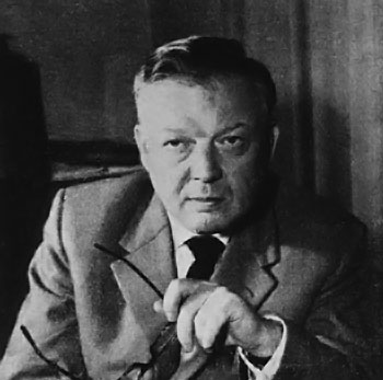
Portrait of Werner Forssmann. Photo taken in 1957.
Source: U.S National Library of Medicine, Images from the History of Medicine Collection Photographer unknown Kindly provided by National Library of Medicine