Award ceremony speech
English
Presentation Speech by Professor Måns Ehrenberg, Member of the Royal Swedish Academy of Sciences; Member of the Nobel Committee for Chemistry, 10 December 2014.
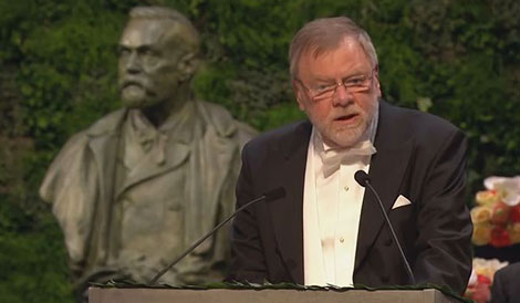
Professor Måns Ehrenberg delivering the Presentation Speech for the 2014 Nobel Prize in Chemistry at the Stockholm Concert Hall.
Your Majesties, Your Royal Highnesses, Ladies and Gentlemen,
This year’s Nobel Prize in Chemistry rewards the development of super-resolved fluorescence microscopy, which has made visible essential details and movements of the molecules of life. This development conquered a physical limit, described by Ernst Abbe in 1873, implying that objects of smaller dimensions than half of the wave length of light cannot be discerned by optical microscopy. Hard as steel seemed Abbe’s law and stern were its prescriptions, but the Norns had a different view, and fate paved the way not for just one, but for two ways to surpass Abbe’s magical limit.
As a young man, Stefan W. Hell thought that one day he would conquer that vexing limit defined by Abbe, but he wasn’t clear about how. He applied for post-doctoral positions to realise his vision, but his request was denied wherever he tried, except in Turku, Finland, where he found a breathing space and valuable time to think. In 1994 he published his theory for how to conquer Abbe’s limit. Two laser beams shine on the structure of interest in a fluorescence microscope. One beam excites the fluorescent molecules in a volume determined by Abbe’s limit. The other beam rapidly brings all the excited molecules, except those in a volume that can be made arbitrarily small, to their ground state before they can emit a photon. When the two beams jointly scan the structure, an image with a resolution far better than Abbe’s limit emerges. In 2000 Hell demonstrated experimentally that his super-resolved fluorescence microscopy works. He called it STimulated Emission Depletion (STED) microscopy. This was the first method to by-pass Abbe’s limit, and now we will turn to the second.
In 1989, William E. Moerner studied light absorption by fluorescent molecules in a crystal matrix at minus 269° C. He then realised that he was the first person in the world to actually observe single, fluorescent electronic dipoles: single molecule spectroscopy was born.
Now a quick jump ahead to 1995. Eric Betzig was tired of university life. He wanted to do something else, but could not stop thinking about how to use single molecule detection and fluorescence microscopy to eliminate Abbe’s damned limit. It was well established that when fluorescent groups that label a biological structure are further apart than Abbe’s limit, they can be localised with unlimited precision. However, with such sparse labelling there would be big information holes in the structure. To solve this problem, Betzig would have to navigate between the Scylla of Abbe’s limit and the Charybdis of the information holes, and he was frustrated. He theoretically described these concepts in 1995, but there was no obvious way to implement them in practice. Betzig left the university world and went into exile, but the basis for a practical way to implement his concepts would soon come from Moerner’s single molecule characterisation of an amazing variant of the green fluorescent protein (GFP).
Moerner discovered in 1997 that a new variant of GFP could be activated from a non-fluorescent to a fluorescent state using light of a wave length that was very different from that of the exciting light. This GFP molecule served as a a molecular lamp that could be turned on and off by an optical switch.
When Betzig finally came back from his exile, the solution to his problem was thus within reach. He developed a method where the biological structure is labelled with optically activatable GFP. Then a weak light pulse activates a small fraction of the GFP molecules, so that all molecules in this fraction are further apart than Abbe’s limit, and thus can be localised with super-precision. Then, yet another small fraction of the GFP molecules are activated so that also they can be super-localised. This is repeated until a large number of such images have been created. Finally, all these images are combined into one super-resolved image with complete structural information. Betzig published his method in 2006 and called it Photo Activated Localisation Microscopy (PALM).
Dr Betzig, Professors Hell and Moerner:
You are being awarded the Nobel Prize in Chemistry for the development of super-resolved fluorescence microscopy. On behalf of the Royal Swedish Academy of Sciences, I wish to convey to you our warmest congratulations and I now ask you to step forward to receive your Nobel Prizes from the hand of His Majesty the King.
Nobel Prizes and laureates
Six prizes were awarded for achievements that have conferred the greatest benefit to humankind. The 14 laureates' work and discoveries range from quantum tunnelling to promoting democratic rights.
See them all presented here.

