Revealing secrets on a tiny scale
The small matter of what everything is made from has long fascinated humankind. Microscopes have had a huge impact on our understanding of everything from the composition of materials to the building blocks of life. The work of a number of Nobel Prize laureates have allowed microscopes to evolve.
A fascinating microbial world
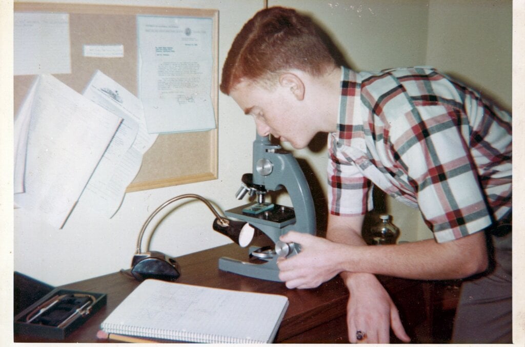
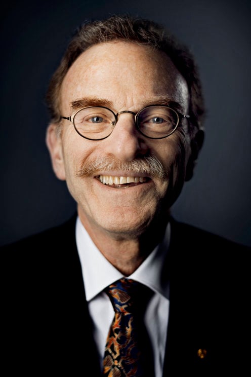
Peering into the plastic eyepiece of his toy microscope to take a closer look at a drop of pond scum, Randy Schekman was transported into a fascinating microbial world. Aged 12, he was determined to save money from odd jobs to buy his first professional microscope. But he couldn’t reach his goal as his mother kept borrowing money from his piggy bank.
“One Saturday I became so upset that, after mowing a neighbour’s lawn, I bicycled to the police station and announced to the desk officer that I wanted to run away from home because my mother took my money and I couldn’t use it to buy a microscope,” he said.
The desperate measures paid off: his parents picked him up from the police station and bought the microscope on their way home. It remained a “treasured possession” throughout Schekman’s school years.
Making the invisible, visible
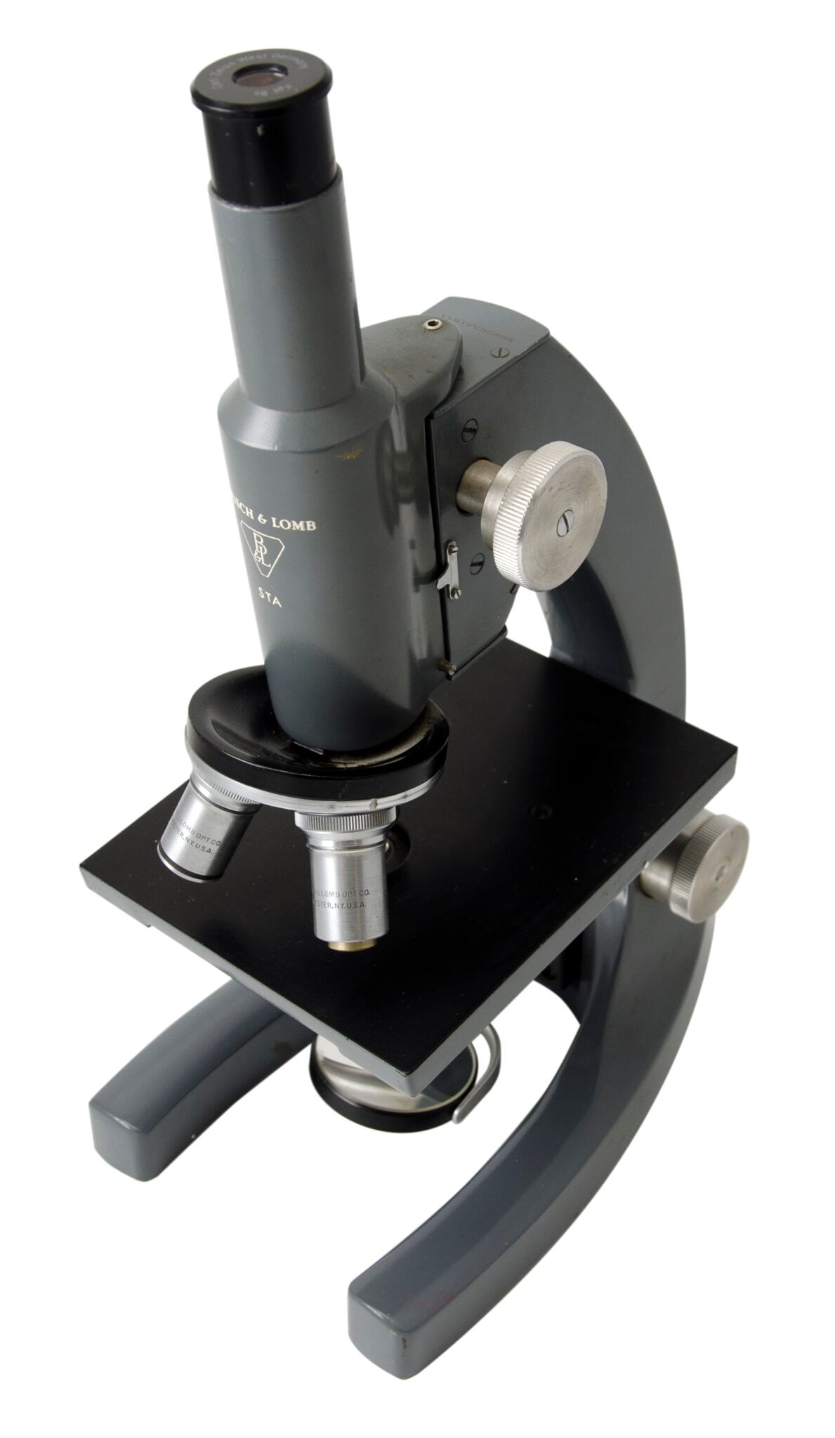
The microscope’s ability to be an extension of the human eye has enabled scientists to make strides in biology and medicine since its invention in the 16th century. Around one century ago, a scientific breakthrough by Richard Zsigmondy led to the development of the ultramicroscope.
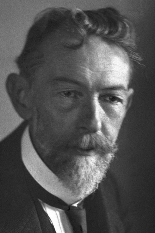
But it was not until Frits Zernike took a fresh look at how refraction gives rise to the image in a microscope that scientists could distinguish transparent specimens such as bacteria and cells without having to cleverly illuminate or stain them.
When light rays pass through transparent materials, such as biological specimens, there is a change in the phase of the light waves – the position of the wave crests in relation to one another – compared to an unimpeded light ray. Our eyes can’t detect this but these phase changes contain important information that can be used to visualise the material they have passed through.
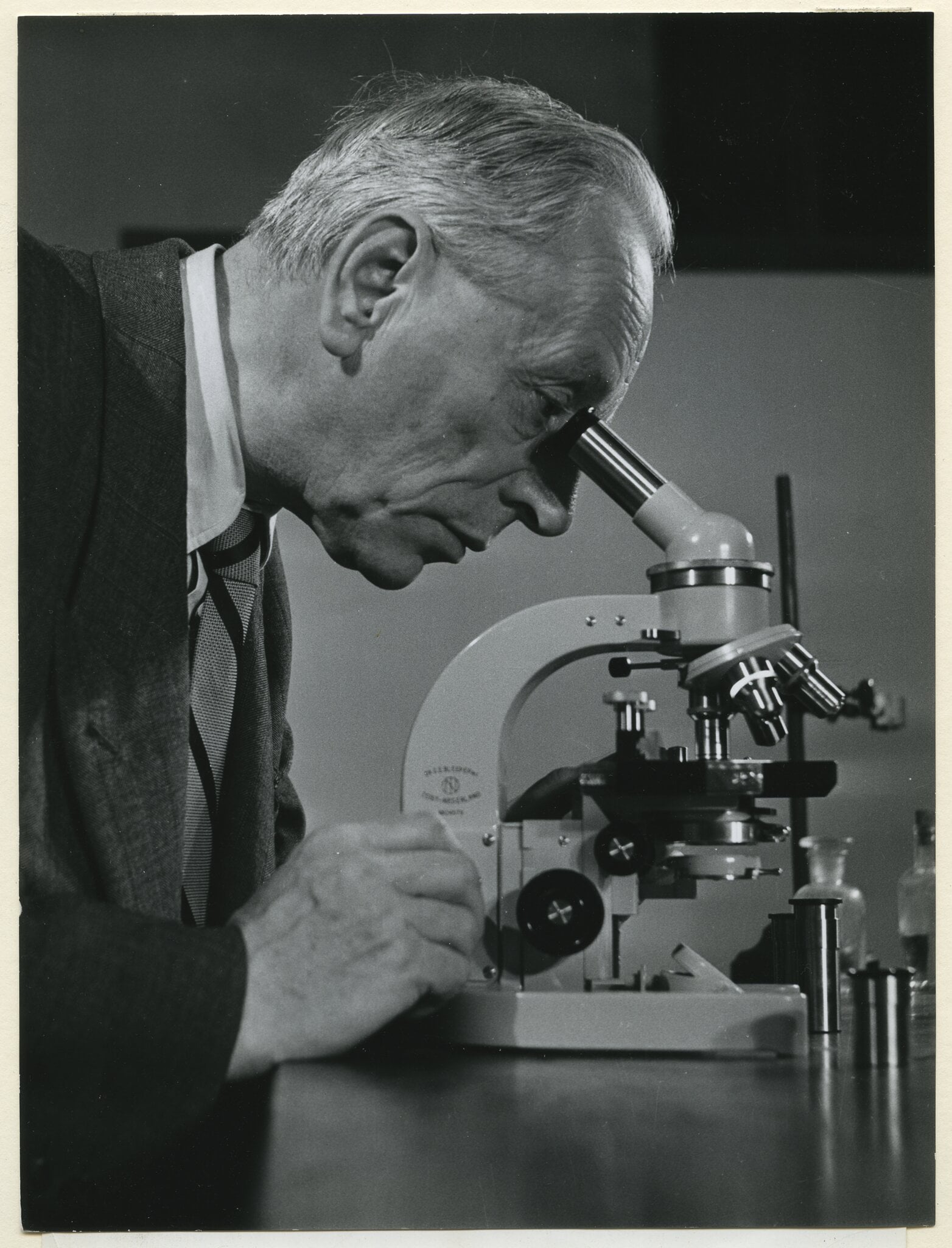
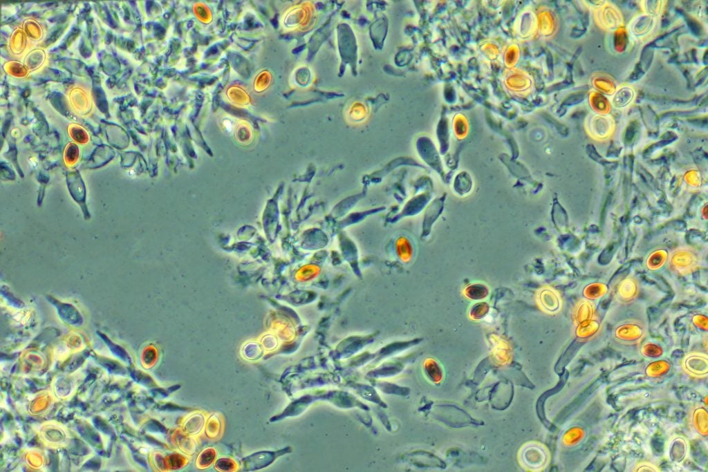
Zernike worked out a way to make this phase change visible, and built the phase contrast microscope in the 1930s. His microscope was capable of enhancing the contrast of unstained, transparent specimens to reveal their inner workings in richer detail, revolutionising biological and medical research.
Despite being told by a major microscope manufacturer that his invention had “little practical value”, Zernike’s work had a massive impact that continues to be felt.
Phase contrast microscopes have enabled strides in biological and medical research to be made, especially in the fields of histology – the study of tissues and organs in the body – and cancer research. They also allow commercial products such as oils, drugs and textiles to be scrutinised.
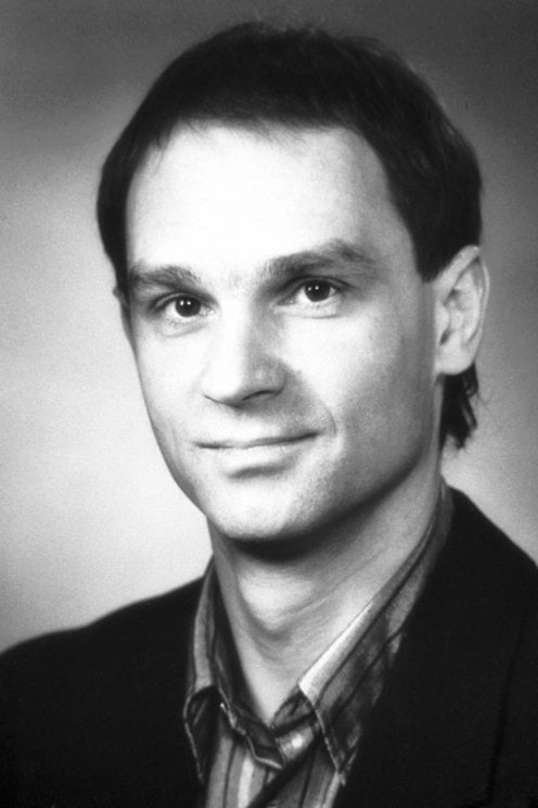
Seeing life at an atomic level
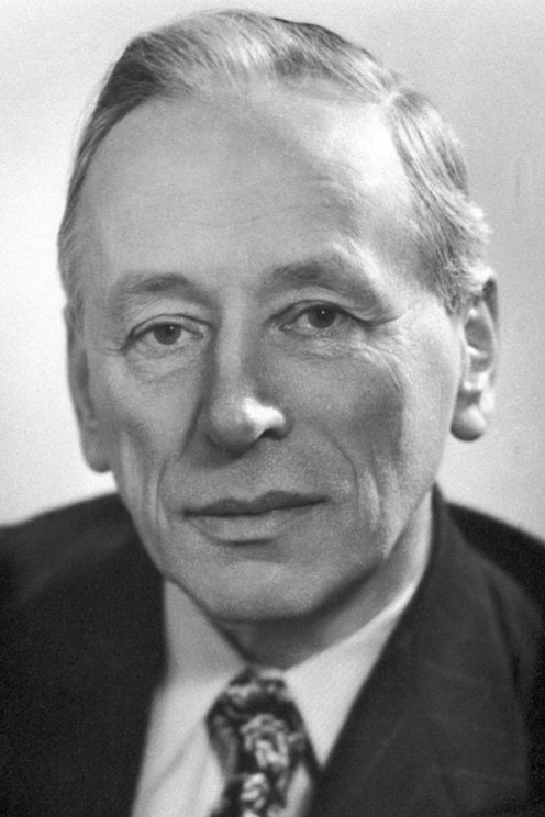
Ernst Ruska also had to overcome criticism, which began at an early age. Yet, Ruska’s conviction and curiosity paid off when, as a young student in 1933, he developed the first electron microscope. By using a magnetic coil as a lens he harnessed electron beams – instead of light – to obtain images of extremely small objects. The resulting image had a resolution greater than that of light microscopes, which was a breakthrough for science.
His invention allowed the smallest objects, such as cell organelles, viruses, or atoms, to be examined.
Ruska helped commercialise his invention, so that mass-produced electron microscopes rapidly found applications within many areas of science.
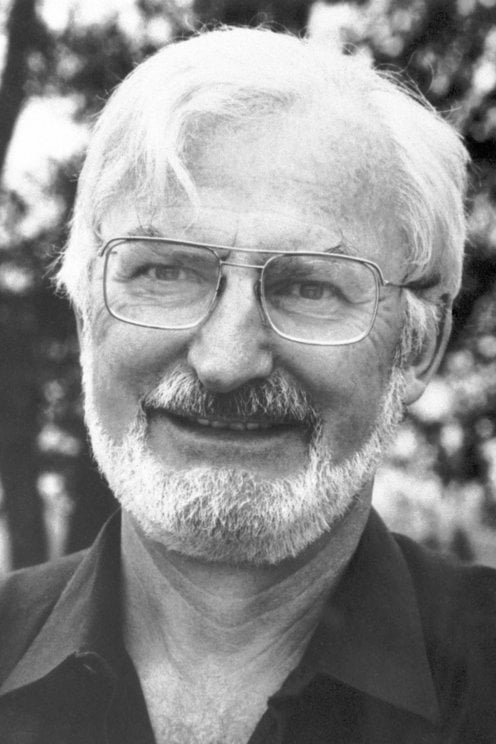
The development of the electron microscope opened up a previously hidden world. Ruska was recognised with the Nobel Prize in Physics 1986, which he shared with Gerd Binnig and Heinrich Rohrer who also turned to electrons to design a new microscope.
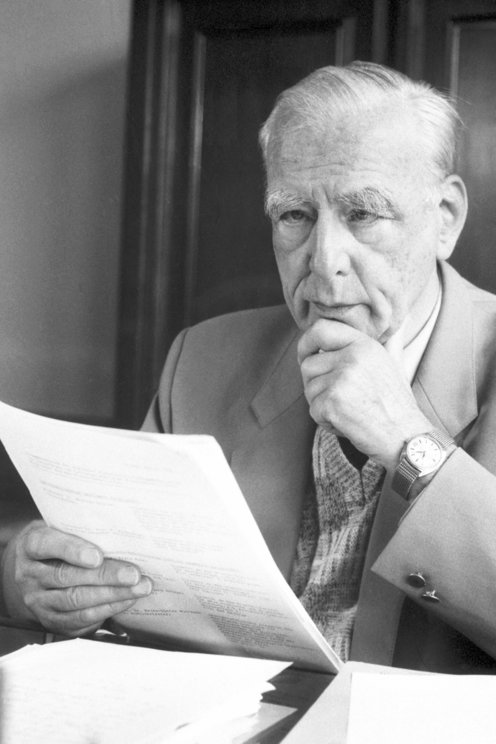
Binnig and Rohrer’s scanning tunnelling microscope (STM) used a single atom-sized stylus to create a topographical map of a surface. They exploited the tunnelling effect, in which a current flows between the stylus and the surface only if they are close enough together, to visualise the surface’s peaks and troughs at the atomic level. Thanks to their advances, crystal surfaces, DNA molecules and viruses could be visualised, opening up new vistas to life around us.
Bringing optical microscopy into the nanodimension
Electron microscopes are incredibly powerful, but they cannot be used to image living cells because the electrons destroy the samples.

For a long time, an alternative – optical microscopy – was held back because scientists presumed that optical microscopes would never obtain a better resolution than half the wavelength of light, and could therefore not be used to visualise tiny objects such viruses and protein molecules in cells.
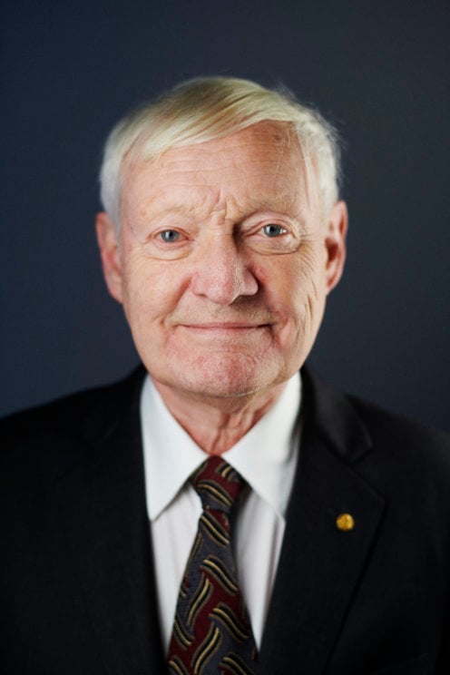
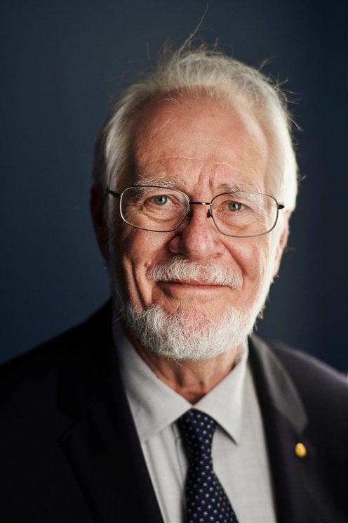
Eric Betzig, Stefan Hell and William Moerner, awarded the Nobel Prize in Chemistry 2014, used fluorescence to bypass this limit. Their work allowed the optical microscope to peer into the nanoworld. Nanoscopy – the ability to see past the optical limit of 200-300nm – revolutionised the field of cell biology. Theoretically, there is no longer any object too small to be studied.
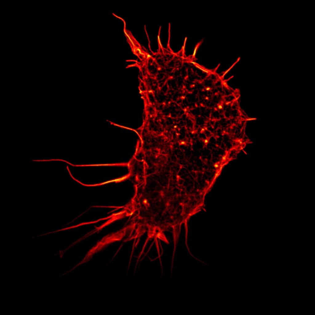
Hell’s technique used fluorescent molecules to image nano-sized parts of a cell. From this he developed the stimulated emission depletion (STED) microscope, which he used at the turn of the millennium to image E. coli bacterium at a resolution never before achieved in an optical microscope.
Betzig and Moerner laid the foundation for a second method; single-molecule microscopy. It relies upon the possibility to turn the fluorescence of individual molecules on and off. Scientists image the same area multiple times, letting just a few interspersed molecules glow each time. Superimposing these images yields a dense super-image resolved at the nanoscale.
Nanoscopy is, among other things, being used to better understand the function of living nerve cells and neuronal circuits. Scientists are working on allowing continuous observation, so they could “film” the workings of “molecular machines” such as proteins, which could be used, for example, to design new drugs.
Bolstering biochemistry
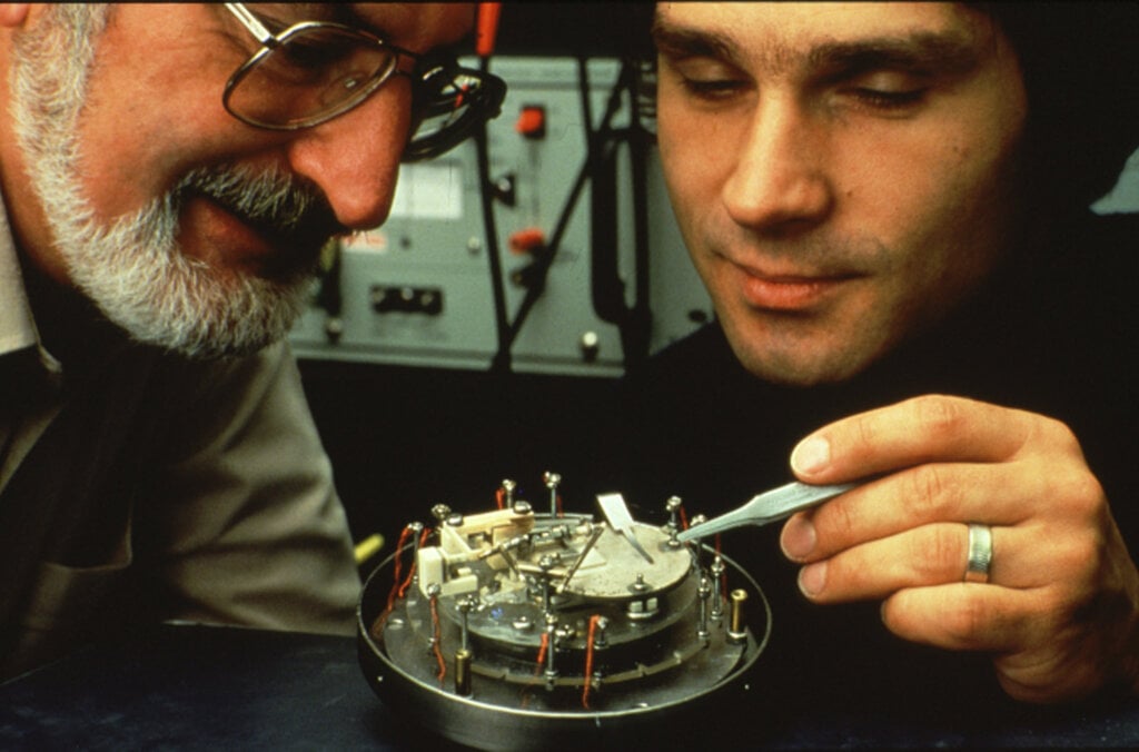
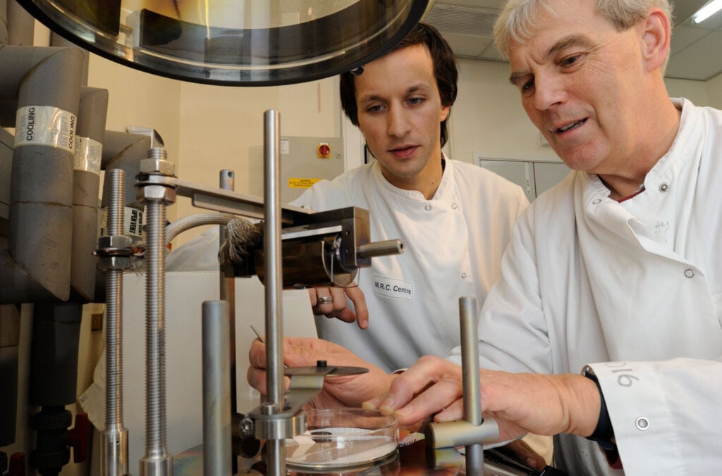
The Nobel Prize in Chemistry 2017 recognised discoveries that contributed to the development of cryo-electron microscopy, which has enabled a new era of biochemistry.
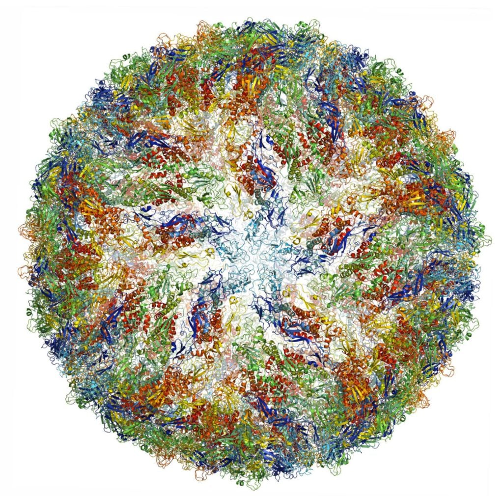
Electron microscopes were only useful for imaging dead objects, because the powerful electron beam destroys biological material. But thanks to the work of Richard Henderson, Joachim Frank and Jacques Dubochet, researchers can now freeze biomolecules mid-movement and portray them at atomic resolution.
Cryo-electron microscopy builds on Henderson and Frank’s research using electron microscopes to visualise biological samples and Dubochet’s method of cooling the water around molecules so rapidly they can be imaged in their ‘natural’ form. Today the technique is being successfully applied to multiple scientific disciplines to reveal molecular insights that would otherwise not be achievable.
In medical research, it is used, for example, to observe the 3D outer shell structures of viruses, uncover the atomic structures of proteins associated with neurodegenerative diseases, and to determine the structure and interactions of proteins that play a significant role in cancer. Better understanding of such mechanisms could lead to the development of more effective treatments.
Focusing on the future
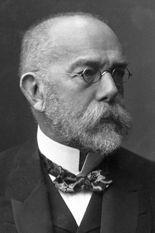
Numerous Nobel Prize laureates have used microscopes to make beneficial breakthroughs for humankind. The founder of modern bacteriology, Robert Koch, reportedly penned a letter to microscope manufacturer Carl Zeiss that read, “a large part of my success I owe to your excellent microscopes,” having discovered the bacilli that caused tuberculosis and cholera, and receiving the Nobel Prize in Physiology or Medicine in 1905.
Microscopes will doubtlessly help future laureates to experience their eureka moments and brilliant breakthroughs. They are more than just instruments, but gateways to other tiny worlds, that continue to enable revolutionary discoveries.
Microscopes and the Nobel Prize
Nobel Prize laureates mentioned in this article


Nobel Prizes and laureates
Six prizes were awarded for achievements that have conferred the greatest benefit to humankind. The 14 laureates' work and discoveries range from quantum tunnelling to promoting democratic rights.
See them all presented here.

