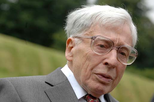
Robert G. Edwards
Photo gallery

1 (of 11)
Mrs Ruth Edwards receiving Robert G. Edwards' Nobel Prize from His Majesty King Carl XVI Gustaf of Sweden at the Stockholm Concert Hall, 10 December 2010.
Copyright © The Nobel Foundation 2010
Photo: Frida Westholm

2 (of 11)
Mrs Ruth Edwards receiving Robert G. Edwards' Nobel Prize at the Stockholm Concert Hall, 10 December 2010.
Copyright © The Nobel Foundation 2010
Photo: AnnaLisa B. Andersson

3 (of 11)
Mrs Ruth Edwards and Professor Martin Hume Johnson showing the Nobel Medal and Diploma of Robert G. Edwards at the Stockholm Concert Hall, 10 December 2010.
Copyright © The Nobel Foundation 2010
Photo: AnnaLisa B. Andersson
4 (of 11)
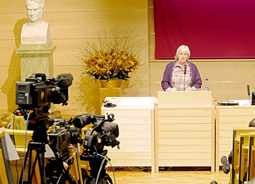
Ruth Edwards (Robert G. Edward's wife) speaking at the Nobel Prize Symposium in Honour of Robert G. Edwards, 7 December 2010.
Copyright © The Nobel Foundation 2010
Photo: Orasisfoto
5 (of 11)
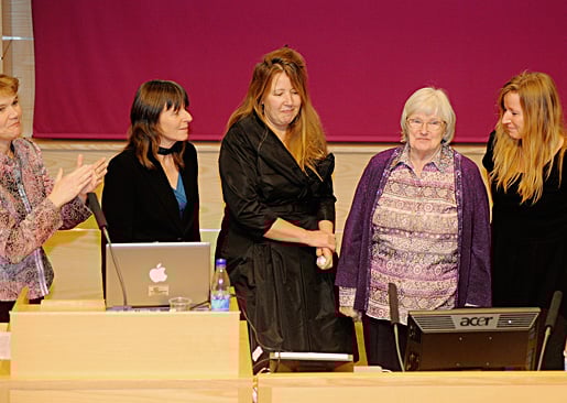
Robert G. Edward's wife, Ruth Edwards, and his daughters, at the Nobel Prize Symposium in Honour of Robert G. Edwards, 7 December 2010.
Copyright © The Nobel Foundation 2010
Photo: Orasisfoto
6 (of 11) Professor Robert Edwards at the Bourn Hall 30th birthday celebrations for first "test tube baby" Louise Brown. Photo taken 12 July 2008.
Photo: Kindly provided by Bourn Hall Clinic.
7 (of 11) Professor Robert Edwards, Lesley Brown, Louise Brown, the world's first "test tube baby" with her son Cameron. Photo taken 12 July 2008.
Photo: Kindly provided by Bourn Hall Clinic.

8 (of 11) Louise Brown, the world's first "test tube baby" with her mother Lesley. Photo taken 9 October, 1978.
Photo: Brian Bould / Daily Mail / Rex Features /IBL Bildbyrå
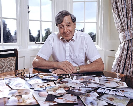
9 (of 11) Professor Robert Edwards at his desk at Bourn Hall Clinic, England. Photo taken in 1989.
Photo: CORBIN O'GRADY STUDIO/ Science Photo Library / IBL Bildbyrå
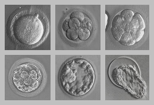
10 (of 11) Human embryos developing in vitro. The photos show a fertilized egg, 8-cell stage, cell adhesion, a compacted morula, a blastocyst and zona hatching.
Photo: Wikipedia (http://en.wikipedia.org/wiki/File:Early_human_embryos.png)
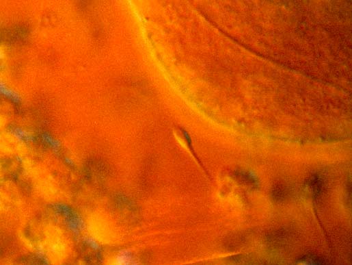
11 (of 11) A light micrograph image showing the encounter between sperm and ovum during in vivo fertilization. A single sperm is seen in centre of the image approaching the circular oocyte. Other competing sperm and cells encompass the corona radiata, which forms a protective halo around the central oocyte, are visible at bottom.
Photo: EDELMANN/SCIENCE PHOTO LIBRARY / IBL Bildbyrå
Nobel Prizes and laureates
Six prizes were awarded for achievements that have conferred the greatest benefit to humankind. The 14 laureates' work and discoveries range from quantum tunnelling to promoting democratic rights.
See them all presented here.

